

A Clinicopathological Investigation of White Matter Hyperintensities and Alzheimer's Disease Neuropathology
- PMID: 29843242
- PMCID: PMC6081639
- DOI: 10.3233/JAD-180017
A Clinicopathological Investigation of White Matter Hyperintensities and Alzheimer's Disease Neuropathology
Abstract
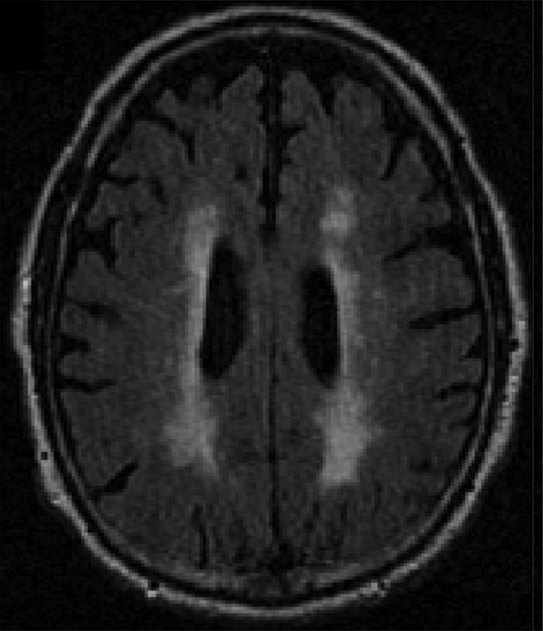
Background: White matter hyperintensities (WMH) on magnetic resonance imaging (MRI) have been postulated to be a core feature of Alzheimer's disease. Clinicopathological studies are needed to elucidate and confirm this possibility.
Objective: This study examined: 1) the association between antemortem WMH and autopsy-confirmed Alzheimer's disease neuropathology (ADNP), 2) the relationship between WMH and dementia in participants with ADNP, and 3) the relationships among cerebrovascular disease, WMH, and ADNP.
Methods: The sample included 82 participants from the National Alzheimer's Coordinating Center's Data Sets who had quantitated volume of WMH from antemortem FLAIR MRI and available neuropathological data. The Clinical Dementia Rating (CDR) scale (from MRI visit) operationalized dementia status. ADNP+ was defined by moderate to frequent neuritic plaques and Braak stage III-VI at autopsy. Cerebrovascular disease neuropathology included infarcts or lacunes, microinfarcts, arteriolosclerosis, atherosclerosis, and cerebral amyloid angiopathy.
Results: 60/82 participants were ADNP+. Greater volume of WMH predicted increased odds for ADNP (p = 0.037). In ADNP+ participants, greater WMH corresponded with increased odds for dementia (CDR≥1; p = 0.038). WMH predicted cerebral amyloid angiopathy, microinfarcts, infarcts, and lacunes (ps < 0.04). ADNP+ participants were more likely to have moderate-severe arteriolosclerosis and cerebral amyloid angiopathy compared to ADNP-participants (ps < 0.04).
Conclusions: This study found a direct association between total volume of WMH and increased odds for having ADNP. In patients with Alzheimer's disease, FLAIR MRI WMH may be able to provide key insight into disease severity and progression. The association between WMH and ADNP may be explained by underlying cerebrovascular disease.
Keywords: Alzheimer’s disease; Alzheimer’s disease neuropathology; cerebrovascular disease; dementia; magnetic resonance imaging; white matter hyperintensities.
Conflict of interest statement
CONFLICT OF INTEREST
RAS is a paid consultant to Eli Lilly (Indianapolis, IN, USA), Avanir Pharmaceuticals (Aliso Viejo, CA), and Biogen (Cambridge, MA, USA). He is a member of the Board of Directors of King-Devick Technologies, Inc. (Chicago, IL, USA), and he receives royalties for published neuropsychological tests from Psychological Assessment Resources, Inc. (Lutz, FL, USA). CD is a paid consultant to Novartis Pharmaceuticals (Basel, Switzerland). For the remaining authors, there are no conflicts of interest to declare.
Figures
References
-
- Johnson KA, Schultz A, Betensky RA, Becker JA, Sepulcre J, Rentz D, Mormino E, Chhatwal J, Amariglio R, Papp K, Marshall G, Albers M, Mauro S, Pepin L, Alverio J, Judge K, Philiossaint M, Shoup T, Yokell D, Dickerson B, Gomez-Isla T, Hyman B, Vasdev N, Sperling R. (2016) Tau positron emission tomographic imaging in aging and early Alzheimer disease. Ann Neurol 79, 110–9. - PMC - PubMed
-
- Kang JH, Korecka M, Figurski MJ, Toledo JB, Blennow K, Zetterberg H, Waligorska T, Brylska M, Fields L, Shah N, Soares H, Dean RA, Vanderstichele H, Petersen RC, Aisen PS, Saykin AJ, Weiner MW, Trojanowski JQ, Shaw LM, Alzheimer’s Disease Neuroimaging I. (2015) The Alzheimer’s Disease Neuroimaging Initiative 2 Biomarker Core: A review of progress and plans. Alzheimers Dement 11, 772–91. - PMC - PubMed
-
- Olsson B, Lautner R, Andreasson U, Ohrfelt A, Portelius E, Bjerke M, Holtta M, Rosen C, Olsson C, Strobel G, Wu E, Dakin K, Petzold M, Blennow K, Zetterberg H. (2016) CSF and blood biomarkers for the diagnosis of Alzheimer’s disease: a systematic review and meta-analysis. Lancet Neurol 15, 673–84. - PubMed
-
- Weiner MW, Veitch DP, Aisen PS, Beckett LA, Cairns NJ, Cedarbaum J, Green RC, Harvey D, Jack CR, Jagust W, Luthman J, Morris JC, Petersen RC, Saykin AJ, Shaw L, Shen L, Schwarz A, Toga AW, Trojanowski JQ, Alzheimer’s Disease Neuroimaging I. (2015) 2014 Update of the Alzheimer’s Disease Neuroimaging Initiative: A review of papers published since its inception. Alzheimers Dement 11, e1–120. - PMC - PubMed
Publication types
MeSH terms
Substances
Grants and funding
- P30 AG013854/AG/NIA NIH HHS/United States
- P50 AG005142/AG/NIA NIH HHS/United States
- U01 NS086659/NS/NINDS NIH HHS/United States
- P30 AG013846/AG/NIA NIH HHS/United States
- K23 NS102399/NS/NINDS NIH HHS/United States
- P50 AG016573/AG/NIA NIH HHS/United States
- P30 AG010161/AG/NIA NIH HHS/United States
- P50 AG025688/AG/NIA NIH HHS/United States
- P50 AG005133/AG/NIA NIH HHS/United States
- R56 NS078337/NS/NINDS NIH HHS/United States
- P30 AG019610/AG/NIA NIH HHS/United States
- P50 AG033514/AG/NIA NIH HHS/United States
- P30 AG010124/AG/NIA NIH HHS/United States
- P50 AG023501/AG/NIA NIH HHS/United States
- P50 AG005131/AG/NIA NIH HHS/United States
- P30 AG010133/AG/NIA NIH HHS/United States
- P50 AG016574/AG/NIA NIH HHS/United States
- P50 AG005146/AG/NIA NIH HHS/United States
- P30 AG035982/AG/NIA NIH HHS/United States
- P50 AG008702/AG/NIA NIH HHS/United States
- U01 AG016976/AG/NIA NIH HHS/United States
- U01 NS093334/NS/NINDS NIH HHS/United States
- P30 AG008051/AG/NIA NIH HHS/United States
- P50 AG005681/AG/NIA NIH HHS/United States
- P50 AG005136/AG/NIA NIH HHS/United States
- P30 AG012300/AG/NIA NIH HHS/United States
- P50 AG016570/AG/NIA NIH HHS/United States
- P50 AG005134/AG/NIA NIH HHS/United States
- P30 AG008017/AG/NIA NIH HHS/United States
- K23 AG046377/AG/NIA NIH HHS/United States
- P50 AG005138/AG/NIA NIH HHS/United States
- R01 NS078337/NS/NINDS NIH HHS/United States
- P30 AG010129/AG/NIA NIH HHS/United States
- P30 AG028383/AG/NIA NIH HHS/United States
- F32 NS096803/NS/NINDS NIH HHS/United States
- UL1 TR001430/TR/NCATS NIH HHS/United States
- RF1 AG054156/AG/NIA NIH HHS/United States
LinkOut - more resources
Full Text Sources
Other Literature Sources
Medical
