Review
. 2018 May 29;7(6):128.
doi: 10.3390/jcm7060128.

Minimally Invasive Limited Ligation Endoluminal-Assisted Revision (MILLER): A Review of the Available Literature and Brief Overview of Alternate Therapies in Dialysis Associated Steal Syndrome

Affiliations
Review

Minimally Invasive Limited Ligation Endoluminal-Assisted Revision (MILLER): A Review of the Available Literature and Brief Overview of Alternate Therapies in Dialysis Associated Steal Syndrome

William W Sheaffer et al. J Clin Med. .

Abstract

Dialysis associated steal syndrome (DASS) is a relatively rare but debilitating complication of arteriovenous fistulas. While mild symptoms can be observed, if severe symptoms are left untreated, DASS can result in ulcerations and limb threatening ischemia. High-flow with resultant heart failure is another documented complication following dialysis access procedures. Historically, open surgical procedures have been the mainstay of therapy for both DASS as well as high-flow. These procedures included ligation, open surgical banding, distal revascularization-interval ligation, revascularization using distal inflow, and proximal invasion of arterial inflow. While effective, open surgical procedures and general anesthesia are preferably avoided in this high-risk population. Minimally invasive limited ligation endoluminal-assisted revision (MILLER) offers both a precise as well as a minimally invasive approach to treating both dialysis associated steal syndrome as well as high-flow with resultant heart failure. MILLER is not ideal for all DASS patients, particularly those with low-flow fistulas. We aim to briefly describe the open surgical therapies as well as review both the technical aspects of the MILLER procedure and the available literature.

Keywords: MILLER; arteriovenous fistula; arteriovenous fistula banding; dialysis associated steal syndrome; high flow.


Conflict of interest statement

The authors declare no conflict of interest.

Figures

Figure 1
Figure 1
Revascularization for steal syndrome of a brachiocephalic arteriovenous fistula (AVF). Brachiocephalic AVF is shown in the center. The darker vessel represents the vein and the lighter vessel represents the artery. DRIL, distal revascularization-interval ligation, is shown on the left. The native artery is ligated distal to the AV anastomosis. Bypass is performed from the native artery proximal to the vascular access take off, to the native artery distal to the site of ligation. The bypass vessel is depicted in the dark color as a saphenous vein graft is commonly used. RUDI, revascularization using distal inflow, is depicted on the right. The original fistula is ligated and the AV anastomosis is relocated distally onto a smaller artery to limit flow to the AVF and also to increase flow through the ulnar artery. Copyright 2018 Mayo Foundation for Medical Education and Research.
Figure 2
Figure 2
Proximalization of arterial inflow (PAI). The existing arteriovenous fistula is dissected and venous outflow is ligated (not shown). A small diameter graft prosthesis is used to deliver proximalized arterial inflow to the new access site. The proximal end of the graft is anastomosed in a side-to-end fashion to the artery and the distal end is anastomosed in an end-to-end fashion to the vein. Copyright 2018 Mayo Foundation for Medical Education and Research.
Figure 3
Figure 3
Banding of a brachiocephalic arteriovenous fistula. The band is placed around the outflow tract of the fistula to create a narrowing which increases resistance to flow and therefore, peripheral perfusion. Copyright 2018 Mayo Foundation for Medical Education and Research.
Figure 4
Figure 4
MILLER (minimally invasive limited ligation endoluminal-assisted revision) steps. (A) A majority of blood flow is diverted into the brachiocephalic arteriovenous fistula (AVF), resulting in less flow to the distal extremity, resulting in steal syndrome. (B) Angioplasty balloon traversing the AVF anastomosis in a retrograde fashion. Incisions are made cranially and caudally to the fistula. (C) Dissection is performed with tunneling of a hemostat beneath the fistula. Monofilament suture is also passed underneath the fistula. The suture is tied firmly around the fistula, but still below the skin, with the endovascular balloon in place as a guide. (D) Post-procedural blood flow is redirected with more blood flowing distally. Copyright 2018 Mayo Foundation for Medical Education and Research.
Figure 5
Figure 5
Angiography during minimally invasive limited ligated endoluminal-assisted revascularization (MILLER) procedure. (A) An inflated 5 mm angioplasty balloon (black arrow) is guided to the proximal arteriovenous graft (AVG) site. A hemostat is used to designate the area for skin incisions. (B) Post-procedure imaging demonstrates a waist in the AVG (black arrow) and distal flow in the brachial artery (red arrow).
Figure 6
Figure 6
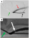
Pre- and post-procedural angiographic images of an arteriovenous graft (AVG) following minimally invasive limited ligation endoluminal-assisted revision. (A) Pre-procedural imaging following brachial artery injection (black arrow) immediately proximal to the anastomosis of the AVG. Contrast is completely diverted into the AVG (red arrow), while no flow is seen distally into the artery (green arrow). (B) Post-procedural imaging shows a newly formed waist (white arrow) in the proximal AVG after the suture has been tied with redistribution of blood flow distally into the brachial artery (green arrow).

References

    1. Saran R., Robinson B., Abbott K.C., Agodoa L.Y., Albertus P., Ayanian J., Balkrishnan R., Bragg-Gresham J., Cao J., Chen J.L., et al. US Renal Data System 2016 Annual Data Report: Epidemiology of Kidney Disease in the United States. Am. J. Kidney Dis. 2017;69:A7–A8. doi: 10.1053/j.ajkd.2016.12.004. - DOI - PMC - PubMed
    1. Malik J., Tuka V., Kasalova Z., Chytilova E., Slavikova M., Clagett P., Davidson I., Dolmatch B., Nichols D., Gallieni M. Understanding the dialysis access steal syndrome. A review of the etiologies, diagnosis, prevention and treatment strategies. J. Vasc. Access. 2008;9:155–166. doi: 10.1177/112972980800900301. - DOI - PubMed
    1. Reddy Y.N.V., Melenovsky V., Redfield M.M., Nishimura R.A., Borlaug B.A. High-Output Heart Failure: A. 15-Year Experience. J. Am. Coll. Cardiol. 2016;68:473–482. doi: 10.1016/j.jacc.2016.05.043. - DOI - PubMed
    1. Reddy Y.N.V., Obokata M., Dean P.G., Melenovsky V., Nath K.A., Borlaug B.A. Long-term cardiovascular changes following creation of arteriovenous fistula in patients with end stage renal disease. Eur. Heart J. 2017;38:1913–1923. doi: 10.1093/eurheartj/ehx045. - DOI - PubMed
    1. Goel N., Miller G.A., Jotwani M.C., Licht J., Schur I., Arnold W.P. Minimally Invasive Limited Ligation Endoluminal-assisted Revision (MILLER) for treatment of dialysis access-associated steal syndrome. Kidney Int. 2006;70:765–770. doi: 10.1038/sj.ki.5001554. - DOI - PubMed

LinkOut - more resources