Rare Fibroepithelial Polyp Extending Along the Ureter: A Case Report
- PMID: 29843497
- PMCID: PMC5981127
- DOI: 10.4274/balkanmedj.2017.1537
Rare Fibroepithelial Polyp Extending Along the Ureter: A Case Report
Abstract
Background: Fibroepithelial polyps of the urinary tract are rare tumors, and their occurrence in the upper urinary tract is highly unusual.
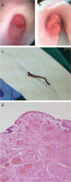
Case report: This study reports a 9-year-old boy who presented to our clinic with complaints of unilateral flank pain and macroscopic hematuria. The direct urinary system graph did not show stone formation; therefore, magnetic resonance urography was performed. This revealed a filling defect in the left proximal ureter. On cystoscopy, a polyp was seen in the orifice of the left ureter, extending along the ureter. The polyp was resected by laser ablation and removed from the ureter. Histopathologic examination revealed a fibroepithelial polyp comprising fibrovascular stroma covered with transitional epithelium.
Conclusion: Although extremely rare, a fibroepithelial polyp should be considered in the differential diagnosis when a young patient presents with flank pain and macroscopic hematuria. Endoscopic procedures may be the treatment of choice for polyps located in the upper ureter.
Keywords: Children; fibroepithelial; polyps; urinary tract.
Conflict of interest statement
Figures
Similar articles
-
Imaging diagnosis and endoscopic treatment for ureteral fibroepithelial polyp prolapsing into the bladder.J Xray Sci Technol. 2013;21(3):393-9. doi: 10.3233/XST-130390. J Xray Sci Technol. 2013. PMID: 24004869
-
[A case of fibroepithelial polyp of the ureter resected ureteroscopically].Hinyokika Kiyo. 2005 Apr;51(4):277-81. Hinyokika Kiyo. 2005. PMID: 15912790 Review. Japanese.
-
Diagnosis and management of ureteral fibroepithelial polyps in children: a new treatment algorithm.J Pediatr Urol. 2015 Feb;11(1):22.e1-6. doi: 10.1016/j.jpurol.2014.08.004. Epub 2014 Aug 28. J Pediatr Urol. 2015. PMID: 25218353 Review.
-
[Ureteral polyp resected with a ureteroscope: report of two cases].Hinyokika Kiyo. 2005 Dec;51(12):809-12. Hinyokika Kiyo. 2005. PMID: 16440729 Japanese.
-
Fibroepithelial ureteral polyp--a case report; endoscopic removal of large ureteral polyp.J Korean Med Sci. 1996 Feb;11(1):80-3. doi: 10.3346/jkms.1996.11.1.80. J Korean Med Sci. 1996. PMID: 8703376 Free PMC article.
Cited by
-
Successful endoscopic treatment using thulium YAG laser for multiple ureteral fibroepithelial polyps in a pediatric patient.IJU Case Rep. 2022 Mar 17;5(3):183-185. doi: 10.1002/iju5.12432. eCollection 2022 May. IJU Case Rep. 2022. PMID: 35509784 Free PMC article.
-
Laparoscopic approach for intermittent hydronephrosis caused by primary ureteral fibroepithelial polyps in children.World J Pediatr Surg. 2021 Mar 11;4(1):e000243. doi: 10.1136/wjps-2020-000243. eCollection 2021. World J Pediatr Surg. 2021. PMID: 36474643 Free PMC article.
-
Fibroepithelial polyps causing obstructive hydronephrosis treated with pyeloplasty: A case report.Urol Case Rep. 2024 Aug 22;56:102828. doi: 10.1016/j.eucr.2024.102828. eCollection 2024 Sep. Urol Case Rep. 2024. PMID: 39257681 Free PMC article.
References
-
- Lam JS, Bingham JB, Gupta M. Endoscopic treatment of fibroepithelial polyps of the renal pelvis and ureter. Urology. 2003;62:810–3. - PubMed
-
- Bhalla RS, Schulsinger DA, Wasnick RJ. Treatment of bilateral fibroepithelial polyps in a child. J Endourol. 2002;16:581–2. - PubMed
-
- Oğuzkurt P, Oz S, Oğuzkurt L, Kayaselçuk F, Tercan F. An unusual cause of complete distal ureteral obstruction: giant fibroepithelial polyp. J Pediatr Surg. 2004;39:1733–4. - PubMed
-
- Güneş M, Keleş MO, Koca OM, Yılmaz Ö, Kaya C. Bilateral üreter obstrüksiyonunun nadir bir sebebi: Fibroepitelyal polip. Marmara Med J. 2010;23:382–5.
-
- Carey RI, Bird VG. Endoscopic management of 10 separate fibroepithelial polyps arising in a single ureter. Urology. 2006;67:413–5. - PubMed
Publication types
MeSH terms
LinkOut - more resources
Full Text Sources
Other Literature Sources


