

Dyke-Davidoff-Masson syndrome: a case report
- PMID: 29843624
- PMCID: PMC5972440
- DOI: 10.1186/s12883-018-1079-3
Dyke-Davidoff-Masson syndrome: a case report
Abstract
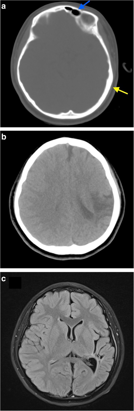
Background: Dyke-Davidoff-Masson syndrome is a rare condition of unknown frequency resulting from brain injury due to a multitude of causes; especially in early life. Characteristics include cerebral hemiatrophy/hypoplasia, contralateral hemiparesis, seizures, and compensatory osseous hypertrophy.
Case presentation: We present a case of a 13-year-old girl who initially presented with headaches, followed by episodic complex-partial seizures; which was controlled via medication. She also had right sided hemiparesis. Computed tomography (CT) showed evidence of left parieto-temporal infarct with cerebral atrophy. Complementary magnetic resonance imaging (MRI) did not reveal additional information. Workup for young stroke was negative. Upon further evaluation by Neuroradiology, features suggesting Dyke-Davidoff-Masson syndrome were confirmed. Patient has been under Neurology follow up since.
Conclusions: Due to its rarity, Dyke-Davidoff-Masson syndrome may easily be missed by the majority of treating clinicians. Knowledge of its features on imaging enables timely and accurate diagnosis - allowing appropriate management.
Keywords: Computed tomography (CT); Dyke-Davidoff-Masson syndrome; Magnetic resonance imaging (MRI).
Conflict of interest statement
Ethics approval and consent to participate
Both authors’ institution does not require ethical approval for publication of a single case report. Written informed consent was obtained from the patient’s next of kin (parents).
Consent for publication
Written informed consent for publication of clinical details and images was obtained from the patient’s next of kin (parents).
Competing interests
The authors declare that they have no competing interests.
Publisher’s Note
Springer Nature remains neutral with regard to jurisdictional claims in published maps and institutional affiliations.
Figures
References
-
- Dyke CG, Davidoff LM, Masson CB. Cerebral hemiatrophy and homolateral hypertrophy of the skull and sinuses. Surg Gynecol Obstet. 1933;57:588–600.
Publication types
MeSH terms
LinkOut - more resources
Full Text Sources
Other Literature Sources
Medical
