Case Reports
. 2018 May 29;18(1):75.
doi: 10.1186/s12883-018-1081-9.

Horizontal gaze palsy with progressive scoliosis: a case report with magnetic resonance tractography and electrophysiological study

Affiliations
Case Reports

Horizontal gaze palsy with progressive scoliosis: a case report with magnetic resonance tractography and electrophysiological study

Chi-Wei Lin et al. BMC Neurol. .

Abstract

Background: Horizontal gaze palsy with progressive scoliosis (HGPPS) is a rare autosomal recessive congenital anomaly characterized by horizontal gaze limitation and progressive scoliosis. We investigated the underlying pathogenesis by incorporating diffusion tensor imaging and an electrophysiological study.

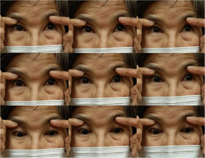
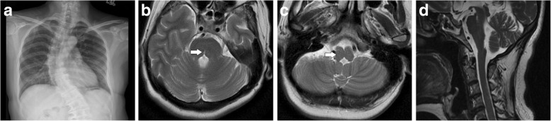
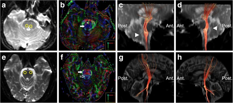
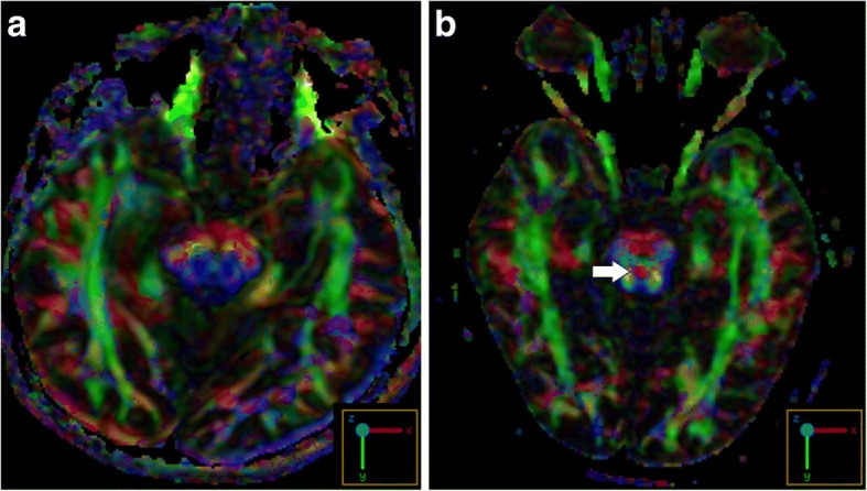
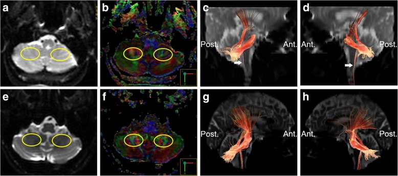
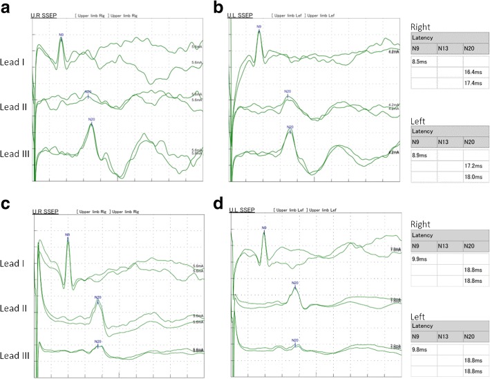
Case presentation: A 55-year-old female patient presented to our clinic due to a chronic history of eye movement limitation since childhood. Her eye problem was followed by a progressive scoliotic change in her torso during junior high school. Neurological examinations revealed remarkable conjugate horizontal but not vertical gaze palsy. Her pupils were isocoric, with a prompt response to light reflex and convergence. Her vision, including visual acuity and field, were normal. No pathological signs of muscle tone, muscle power, deep tendon reflex or coordination were revealed. There was no associated family history, and no diseases involving other systems were noted. On reviewing her past medical history, X-rays revealed scoliotic changes of her thoracic and lumbar spine. Brain magnetic resonance imaging showed a midline cleavage at the tegmentum (split pons sign) and butterfly configuration of the medulla, consistent with HGPPS. Color-coded diffusion tensor imaging in our patient revealed absence of decussation of the superior cerebellar peduncle. In tractography, the pontocerebellar tracts and fibers within the inferior cerebellar peduncle, deemed to be primarily dorsal spinocerebellar and vestibulocerebellar tracts, appeared to be agenetic. The tegmentum was compromised secondary to dorsal displacement of the corticospinal tracts. Of note, the bilateral corticospinal tracts remained uncrossed at the level presumed to be the pyramidal decussation. A somatosensory evoked potential study also revealed predominantly ipsilateral cortical sensory responses.

Conclusions: Our study confirmed that a compromised tegmentum secondary to dorsal displacement of the corticospinal tracts and poorly-developed afferent fibers within the pontocerebellar tracts and inferior cerebellar peduncle to be the main neuroanatomical anomalies responsible for the clinical presentations of HGPPS. In addition, the uncrossed nature of the majority of pyramidal and proprioceptive sensory systems was confirmed.

Keywords: Diffusion tensor imaging; Horizontal gaze palsy with progressive scoliosis; Magnetic resonance imaging; Somatosensory evoked potential; Split pons sign; Tractography.


Conflict of interest statement

Ethics approval and consent to participate

The authors declare that ethics approval was not required for this case report.

Consent for publication

Written informed consent was obtained from the patient and the healthy control for publication in this Case Report and any accompanying images. A copy of the written consent form is available for review by the Editor of this journal.

Competing interests

The authors declare that they have no competing interests.

Publisher’s Note

Springer Nature remains neutral with regard to jurisdictional claims in published maps and institutional affiliations.

Figures

Fig. 1
Fig. 1
Cardinal directions of gaze in this patient. There was a limited range of voluntary ductions in horizontal directions. Vertical gazing was normal
Fig. 2
Fig. 2
Main conventional imaging findings of this patient with horizontal gaze palsy with progressive scoliosis. a Chest X-ray showing S-shaped scoliosis of the thoracolumbar spine. b Axial T2-weighted brain magnetic resonance imaging showing a midline cleavage at the dorsal aspect of the pons (split pons sign) (arrow). c Butterfly configuration of the medulla (arrow). d Atrophic change of the pons in sagittal view
Fig. 3
Fig. 3
Diffusion tensor imaging showing an anatomical anomaly within the pons. a, e Selection of the seed regions of interest (35 mm2) in axial T2 weighted imaging. b, f Color-coded diffusion tensor imaging showing dorsal displacement of bilateral corticospinal tracts (solid arrows), malposition of the transverse pontine fibers (asterisk), and agenesis of the medial longitudinal fasciculus (hollow arrow in the healthy control). Tractography on the right (c, g) and left (d, h) side suggested agenesis of the pontocerebellar tracts (arrow heads) in the patients compared to the healthy control. Upper row: the patient; lower row: the age and sex-matched healthy control. Ant.: Anterior side; Post.: Posterior side
Fig. 4
Fig. 4
Color-coded diffusion tensor imaging showing absence of red dot in this patient (a) representing decussation of the superior cerebellar peduncle normally seen in the healthy control (arrow) (b)
Fig. 5
Fig. 5
Tractography of the patient showing the uncrossed bilateral corticospinal tracts (a, b, d, e the seed regions selection; c, f the uncrossed bilateral corticospinal tracts). No crossed fibers were identified on placing the seed regions of the spinal cord contralateral to those within the pons
Fig. 6
Fig. 6
Tractography showing atrophic fibers within the inferior cerebellar peduncle (arrows). a, e Selection of the seed regions of interest (350 mm2) in axial T2 weighted imaging at approximately the same level. b, f Color-coded diffusion tensor imaging. c, g Tractography on the right-side. d, h Tractography on the left side. Upper row: the patient; lower row: the age and sex-matched healthy control. Ant.: Anterior side; Post.: Posterior side
Fig. 7
Fig. 7
Somatosensory evoked potential studies comparing the patient and healthy control. Predominant signals of the cortex were detected ipsilateral to the stimulus in the patient (a, b), in contrast to predominant signals of the cortex contralateral to the stimulus in the healthy control (c, d). a, c the right-side stimulus. b, d the left-side stimulus. Lead I: ipsilateral Erb point, Lead II: contralateral cortex, Lead III: ipsilateral cortex

References

    1. Rossi A, Catala M, Biancheri R, Di Comite R, Tortori-Donati P. MR imaging of brain-stem hypoplasia in horizontal gaze palsy with progressive scoliosis. AJNR Am J Neuroradiol. 2004;25:1046–1048. - PMC - PubMed
    1. dos Santos AV, Matias S, Saraiva P, Goulao A. MR imaging features of brain stem hypoplasia in familial horizontal gaze palsy and scoliosis. AJNR Am J Neuroradiol. 2006;27:1382–1383. - PMC - PubMed
    1. Jen JC, Chan WM, Bosley TM, Wan J, Carr JR, Rub U, et al. Mutations in a human ROBO gene disrupt hindbrain axon pathway crossing and morphogenesis. Science. 2004;304:1509–1513. doi: 10.1126/science.1096437. - DOI - PMC - PubMed
    1. Pieh C, Lengyel D, Neff A, Fretz C, Gottlob I. Brainstem hypoplasia in familial horizontal gaze palsy and scoliosis. Neurology. 2002;59:462–463. doi: 10.1212/WNL.59.3.462. - DOI - PubMed
    1. Otaduy MC, Leite Cda C, Nagae LM, Pinho Mda C, Bueno C, Reed UC, et al. Further diffusion tensor imaging contribution in horizontal gaze palsy and progressive scoliosis. Arq Neuropsiquiatr. 2009;67:1054–1056. doi: 10.1590/S0004-282X2009000600017. - DOI - PubMed

Publication types

Supplementary concepts