. 2018 May 29;18(1):91.
doi: 10.1186/s12890-018-0652-2.

Stability of gene expression by primary bronchial epithelial cells over increasing passage number

Affiliations

Stability of gene expression by primary bronchial epithelial cells over increasing passage number

Stephen R Reeves et al. BMC Pulm Med. .

Abstract

Background: An increasing number of studies using primary human bronchial epithelial cells (BECs) have reported intrinsic differences in the expression of several genes between cells from asthmatic and non-asthmatic donors. The stability of gene expression by primary BECs with increasing cell passage number has not been well characterized.

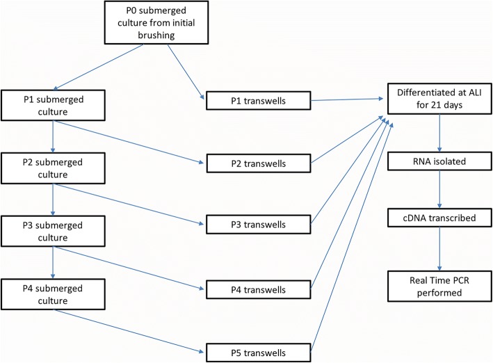
Methods: To determine if expression by primary BECs from asthmatic and non-asthmatic children of selected genes associated with airway remodeling, innate immune response, immunomodulatory factors, and markers of differentiated airway epithelium, are stable over increasing cell passage number, we studied gene expression patterns in passages 1, 2, 3, 4, and 5 BECs from asthmatic (n = 6) and healthy (n = 6) subjects that were differentiated at an air-liquid interface. RNA was harvested from BECs and RT-PCR was performed for TGFβ1, TGFβ2, activin A, FSTL3, MUC5AC, TSLP, IL-33, CXCL10, IFIH1, p63, KT5, TUBB4A, TJP1, OCLN, and FOXJ1.

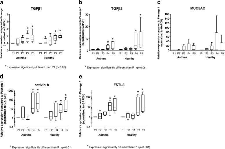
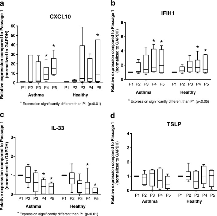
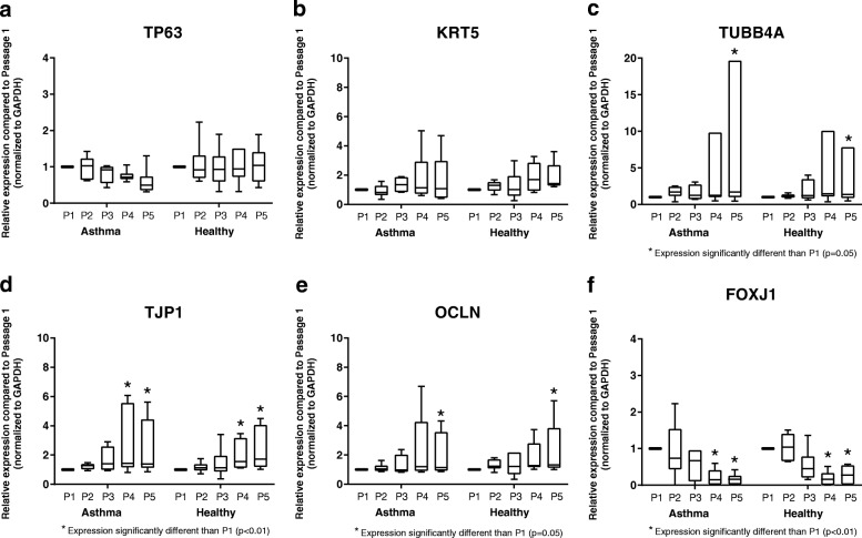
Results: Expression of TGFβ1, TGFβ2, activin A, FSTL3, MUC5AC, CXCL10, IFIH1, p63, KT5, TUBB4A, TJP1, OCLN, and FOXJ1 by primary BECs from asthmatic and healthy children was stable with no significant differences between passages 1, 2 and 3; however, gene expression at cell passages 4 and 5 was significantly greater and more variable compared to passage 1 BECs for many of these genes. IL-33 and FOXJ1 expression was also stable between passages 1 through 3, however, expression at passages 4 and 5 was significantly lower than by passage 1 BECs. TSLP, p63, and KRT5 expression was stable across BEC passages 1 through 5 for both asthmatic and healthy BECs.

Conclusions: These observations illustrate the importance of using BECs from passage ≤3 when studying gene expression by asthmatic and non-asthmatic primary BECs and characterizing the expression pattern across increasing cell passage number for each new gene studied, as beyond passage 3 genes expressed by primary BECs appear to less accurately model in vivo airway epithelial gene expression.

Keywords: Airway remodeling; Asthma; Children; Epithelial cells.


Conflict of interest statement

Ethics approval and consent to participate

Written consent was obtained from parents of subjects and assent was obtained for children ≥ age 10 years to participate in this. The work presented in this study was approved by the Seattle Children’s Hospital Institutional Review Board.

Competing interests

The authors declare that they have no competing interests, financial or non-financial.

Publisher’s Note

Springer Nature remains neutral with regard to jurisdictional claims in published maps and institutional affiliations.

Figures

Fig. 1
Fig. 1
Schematic depicting experimental design for passaging primary bronchial epithelial cells over 5 passages in ex vivo cell culture for gene expression studies
Fig. 2
Fig. 2
Expression of genes related to airway remodeling by primary BECs. Expression of TGFβ1 (a), TGFβ2 (b), MUC5AC (c), activin A (d), and FSTL3 (e) by BECs at P1 (n = 6 asthma donors, n = 6 healthy donors), P2 (n = 6 asthma donors, n = 6 healthy donors), P3 (n = 4 asthma donors, n = 6 healthy donors), P4 (n = 6 asthma donors, n = 6 healthy donors), and P5 (n = 6 asthma donors, n = 6 healthy donors) are presented as box-and-whisker plots which depict the interquartile range and median (the ends of each box represent the upper and lower quartiles, error bars represent the maximum and minimum, and the horizontal line within the box represents the median). To compare expression of genes at P2-P5 to expression at P1, and to compare patterns of gene expression between asthmatic and healthy donors, ordinary two-way ANOVA with Dunnett’s multiple comparisons test was used for normally distributed data, and Kruskal-Wallis ANOVA with Dunn’s multiple comparisons test was used for non-normally distributed data
Fig. 3
Fig. 3
Expression of innate immunity and immunomodulatory genes by primary BECs. Expression of CXCL10 (a), IFIH1 (b), IL-33 (c), and TSLP (d) by BECs at P1 (n = 6 asthma donors, n = 6 healthy donors), P2 (n = 6 asthma donors, n = 6 healthy donors), P3 (n = 4 asthma donors, n = 6 healthy donors), P4 (n = 6 asthma donors, n = 6 healthy donors), and P5 (n = 6 asthma donors, n = 6 healthy donors) are presented as box-and-whisker plots which depict the interquartile range and median (the ends of each box represent the upper and lower quartiles, error bars represent the maximum and minimum, and the horizontal line within the box represents the median). To compare expression of genes at P2-P5 to expression at P1, and to compare patterns of gene expression between asthmatic and healthy donors, ordinary two-way ANOVA with Dunnett’s multiple comparisons test was used for normally distributed data, and Kruskal-Wallis ANOVA with Dunn’s multiple comparisons test was used for non-normally distributed data
Fig. 4
Fig. 4
Expression of genes associated with airway epithelial basal cells, ciliogenesis, and epithelial tight junctions. Expression of TP63 (a), KRT5 (b), TUBB4A (c), TJP1 (d), OCLN (e), and FOXJ1 (f) by BECs at P1 (n = 6 asthma donors, n = 6 healthy donors), P2 (n = 6 asthma donors, n = 6 healthy donors), P3 (n = 4 asthma donors, n = 6 healthy donors), P4 (n = 6 asthma donors, n = 6 healthy donors), and P5 (n = 6 asthma donors, n = 6 healthy donors) are presented as box-and-whisker plots which depict the interquartile range and median (the ends of each box represent the upper and lower quartiles, error bars represent the maximum and minimum, and the horizontal line within the box represents the median). To compare expression of genes at P2-P5 to expression at P1, and to compare patterns of gene expression between asthmatic and healthy donors, ordinary two-way ANOVA with Dunnett’s multiple comparisons test was used for normally distributed data, and Kruskal-Wallis ANOVA with Dunn’s multiple comparisons test was used for non-normally distributed data

References

    1. The global asthma report 2014. Auckland: Global Asthma Network; 2014. www.globalasthmanetwork.org.
    1. Holgate ST. The sentinel role of the airway epithelium in asthma pathogenesis. Immunol Rev. 2011;242:205–219. doi: 10.1111/j.1600-065X.2011.01030.x. - DOI - PubMed
    1. Holtzman MJ, Byers DE, Alexander-Brett J, Wang X. The role of airway epithelial cells and innate immune cells in chronic respiratory disease. Nat Rev Immunol. 2014;14:686–698. doi: 10.1038/nri3739. - DOI - PMC - PubMed
    1. McLellan K, Shields M, Power U, Turner S. Primary airway epithelial cell culture and asthma in children-lessons learnt and yet to come. Pediatr Pulmonol. 2015;50:1393–1405. doi: 10.1002/ppul.23249. - DOI - PubMed
    1. Stewart CE, Torr EE, Mohd Jamili NH, Bosquillon C, Sayers I. Evaluation of differentiated human bronchial epithelial cell culture systems for asthma research. J Allergy (Cairo) 2012;2012:943982. - PMC - PubMed

MeSH terms

Substances

LinkOut - more resources