. 2018 May 29;19(1):104.
doi: 10.1186/s12931-018-0802-3.

Therapeutic effects of adipose-tissue-derived mesenchymal stromal cells and their extracellular vesicles in experimental silicosis

Affiliations

Therapeutic effects of adipose-tissue-derived mesenchymal stromal cells and their extracellular vesicles in experimental silicosis

Elga Bandeira et al. Respir Res. .

Abstract

Background: Silicosis is an occupational disease that affects workers who inhale silica particles, leading to extensive lung fibrosis and ultimately causing respiratory failure. Mesenchymal stromal cells (MSCs) have been shown to exert therapeutic effects in lung diseases and represent an alternative treatment for silicosis. Recently, it has been suggested that similar effects can be achieved by the therapeutic use of extracellular vesicles (EVs) obtained from MSCs. The aim of this study was to investigate the effects of adipose-tissue-derived MSCs (AD-MSCs) or their EVs in a model of silicosis.

Methods: Silicosis was induced by intratracheal instillation of silica in C57BL/6 mice. After the onset of disease, animals received saline, AD-MSCs, or EVs, intratracheally.

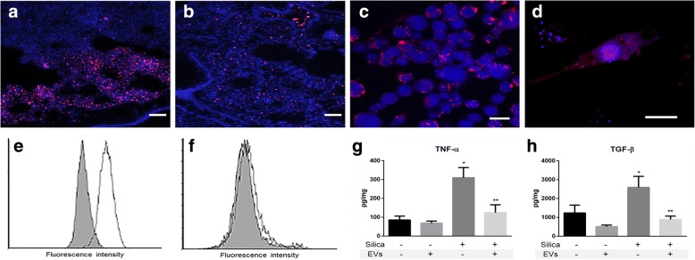
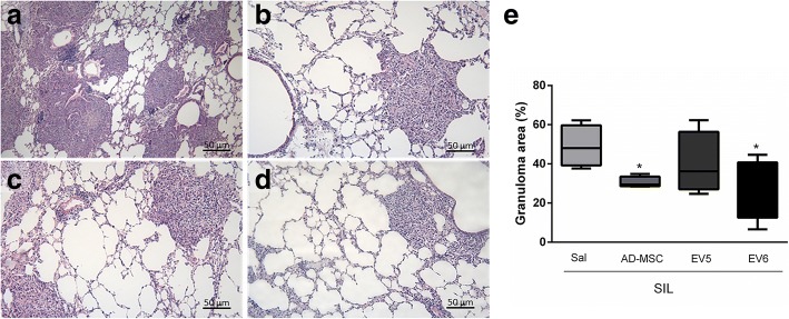
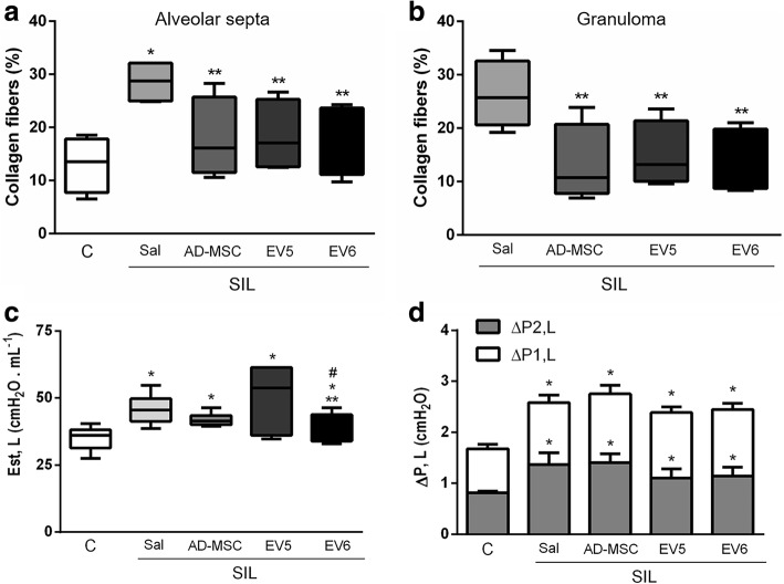
Results: At day 30, AD-MSCs and EVs led to a reduction in collagen fiber content, size of granuloma, and in the number of macrophages inside granuloma and in the alveolar septa. In addition, the expression levels of interleukin 1β and transforming growth factor beta in the lungs were decreased. Higher dose of EVs also reduced lung static elastance when compared with the untreated silicosis group.

Conclusions: Both AD-MSCs and EVs, locally delivered, ameliorated fibrosis and inflammation, but dose-enhanced EVs yielded better therapeutic outcomes in this model of silicosis.

Keywords: Extracellular vesicles; Fibrosis; Inflammation; Mesenchymal stromal cells; Silicosis.


Conflict of interest statement

Ethics approval

This study was approved by the Health Sciences Ethics Committee of the Federal University of Rio de Janeiro (CEUA 188/13). All animals received humane care in compliance with the principles of laboratory animal care formulated by the National Society of Medical Research and the Guide for the Care and Use of Laboratory Animals prepared by the National Academy of Sciences, USA.

Competing interests

The authors declare that they have no competing interests.

Publisher’s Note

Springer Nature remains neutral with regard to jurisdictional claims in published maps and institutional affiliations.

Figures

Fig. 1
Fig. 1
Ultrastructural analysis of the AD-MSC and EV fraction and nanoparticle tracking analysis of the EV fraction. a, b Transmission electron microscopy demonstrates the presence of bulges on the cell membrane, indicating possible microvesicles in AD-MSCs. c Scanning electron microscopy technique of sequential centrifugation-enriched EV fractions of AD-MSC supernatant shows spherical structures ranging from 50 to 100 nm in diameter and the presence of agglomerates. d Size distribution (diameter) of EVs obtained by nanoparticle tracking analysis. Representation of the average from six different samples, with six different videos taken per sample. Curves indicate two Gaussian fittings, and numbers indicate diameter modes
Fig. 2
Fig. 2
Microscopy images showing (a) EV distribution (red) in the lung parenchyma immediately after local instillation; (b) EV distribution (red) in the lung parenchyma 30 min after local instillation; EV uptake (red) by (c) MH-S (alveolar macrophages) × 200 and (d) primary lung fibroblast (× 400, scale bars represent 20 μm). e Flow cytometry representative results of EV uptake (Vybrant DiI staining) by MH-S (alveolar macrophages) after incubation for 24 h. f Flow cytometry representative results of EV uptake (Vybrant DiI staining) by lung primary fibroblasts after incubation for 24 h. Gray areas indicate negative controls. TNF-α (g) and TGF-β (h) concentration (per total amount of protein) in the conditioned medium of cultured macrophages stimulated with silica with or without addition of EVs; bars indicate means ± SEM, n = 4, *Significantly different from unstimulated group. **Significantly different from stimulated group without EVs, p < 0.05
Fig. 3
Fig. 3
Representative photomicrographs of the lung parenchyma of animals from the silica group (a) and the SIL group treated with cells (b) or EVs in two different doses: EV5 (c) and EV6 (d). Slices were stained with hematoxylin–eosin staining. Arrows indicate granulomas, 200×. e Fraction of area of granuloma in the lungs, quantified by analysis of photomicrographs of 5 animals per group, represented as a percentage of the total area of lung tissue. *Significantly different from SIL-Sal, p < 0.05
Fig. 4
Fig. 4
Percentage of F4/80 positive cells (macrophages) in alveolar septa (a) and inside the granulomatous tissue (b). Box plots are representations (min to max) of 20 random field measurements from 5 animals per group. *Significantly different from control group (labeled C). **Significantly different from SIL-Sal group (p < 0.05)
Fig. 5
Fig. 5
Relative measurement of (a) IL-1β, (b) TGF-β, (c) TNFα, (d) IL-6 in the lung tissue. Values were normalized by control levels. *Significantly different from control group (labeled C). **Significantly different from SIL-Sal group (p < 0.05)
Fig. 6
Fig. 6
Impact of silica and treatments with MSCs or EVs in lung remodeling and lung mechanics. a, b Box plot representation of collagen fiber content (min to max) in the alveolar septa (a) or inside the granuloma (b). Values indicate measurements from 10 random fields of 5 animals per group *Significantly different from control group (labeled C). **Significantly different from SIL-Sal group (p < 0.05). c Static lung elastance values in box plot representation (min to max). d Stacked bars represent viscoelastic (ΔP2) and resistive (ΔP1) pressures. Fifteen measurements were taken from each animal, and 6–10 animals were analyzed per group. *Significantly different from control group (labeled C). **Significantly different from SIL-Sal group (p < 0.05)
Fig. 7
Fig. 7
Mean interquartile range, median, and concentration of vesicles (bars/symbols represent means ± SD of 4–6 independent experiments). a Concentration of vesicles per cell normalized by the initial values of freshly obtained vesicles. b Freshly obtained conditioned medium versus thawed medium after 3 weeks of storage at − 80 °C were used to isolate EVs. c Each sample of EVs was measured before and after aerosolization with a microsprayer syringe

References

    1. Leung CC, Yu ITS, Chen W. Silicosis. Lancet. 2012;379:2008–2018. doi: 10.1016/S0140-6736(12)60235-9. - DOI - PubMed
    1. Banks DE, Houston FS, Jindal SK. Can we alter the natural history of silicosis? Chest. 2015;148:57–56. doi: 10.1378/chest.15-0591. - DOI - PubMed
    1. Zhao MM, Cui JZ, Cui Y, Li R, Tian YX, Song SX, et al. Therapeutic effect of exogenous bone marrow-derived mesenchymal stem cell transplantation on silicosis via paracrine mechanisms in rats. Mol Med Rep. 2013;8:741–746. doi: 10.3892/mmr.2013.1580. - DOI - PubMed
    1. Sueblinvong V, Weiss DJ. Stem cells and cell therapy approaches in lung biology and diseases. Transl Res. 2010;156:188–205. doi: 10.1016/j.trsl.2010.06.007. - DOI - PMC - PubMed
    1. Weiss DJ. Stem cells, cell therapies, and bioengineering in lung biology and diseases. Comprehensive review of the recent literature 2010-2012. Ann Am Thorac Soc. 2013;10:S45–S97. doi: 10.1513/AnnalsATS.201304-090AW. - DOI - PMC - PubMed

Publication types

Substances

LinkOut - more resources