. 2018 May 29;11(1):70.
doi: 10.1186/s13045-018-0618-0.

miR-519a enhances chemosensitivity and promotes autophagy in glioblastoma by targeting STAT3/Bcl2 signaling pathway

Affiliations

miR-519a enhances chemosensitivity and promotes autophagy in glioblastoma by targeting STAT3/Bcl2 signaling pathway

Hong Li et al. J Hematol Oncol. .

Abstract

Background: Chemoresistance to temozolomide (TMZ) is a major challenge in the treatment of glioblastoma (GBM). We previously found that miR-519a functions as a tumor suppressor in glioma by targeting the signal transducer and activator of transcription 3 (STAT3)-mediated autophagy oncogenic pathway. Here, we investigated the effects of miR-519a on TMZ chemosensitivity and autophagy in GBM cells. Furthermore, the underlying molecular mechanisms and signaling pathways were explored.

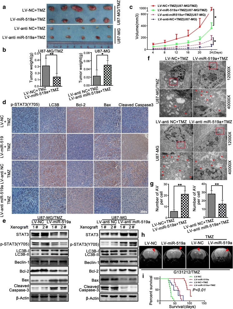
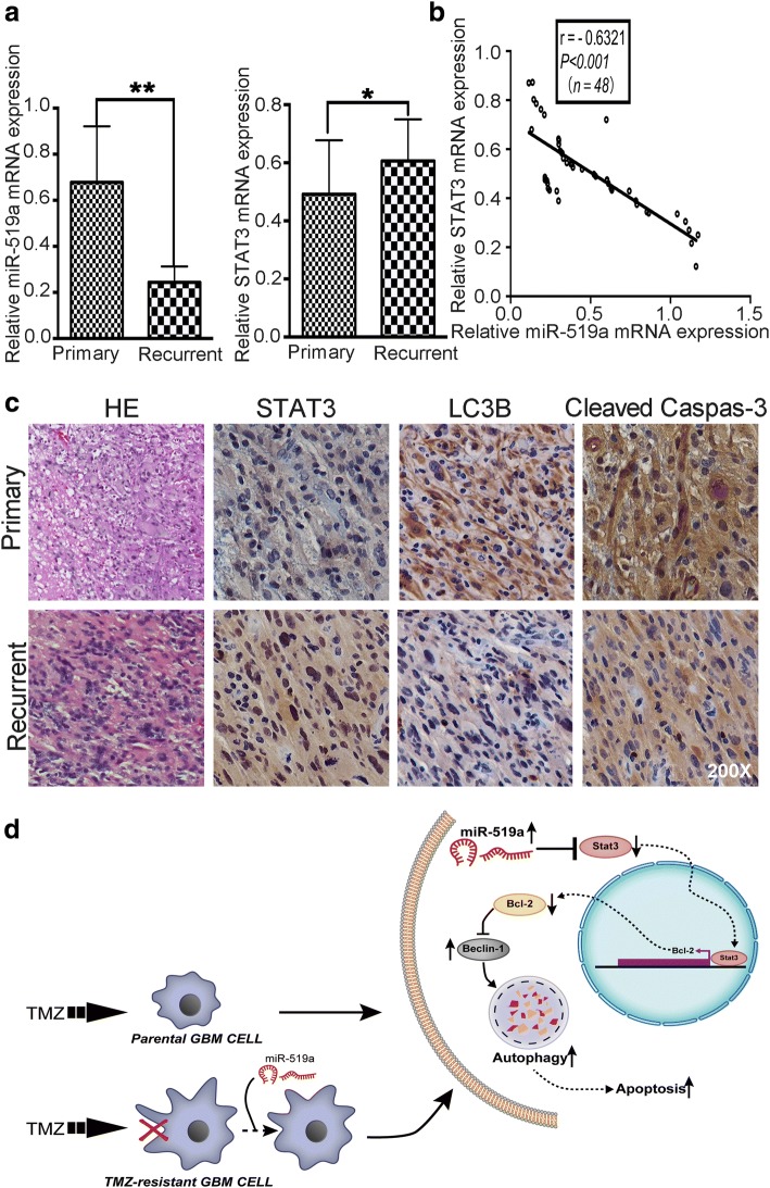
Methods: In the present study, two stable TMZ-resistant GBM cell lines were successfully generated by exposure of parental cells to a gradually increasing TMZ concentration. After transfecting U87-MG/TMZ and U87-MG cells with miR-519a mimic or inhibitor, a series of biochemical assays such as MTT, apoptosis, and colony formation were performed to determine the chemosensitive response to TMZ. The autophagy levels in GBM cells were detected by transmission electron microscopy, LC3B protein immunofluorescence, and Western blotting analysis. Stable knockdown and overexpression of miR-519a in GBM cells were established using lentivirus. A xenograft nude mouse model and in situ brain model were used to examine the in vivo effects of miR-519a. Tumor tissue samples were collected from 48 patients with GBM and were used to assess the relationship between miR-519a and STAT3 expression.

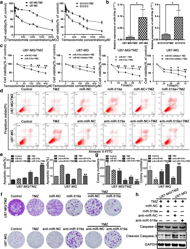
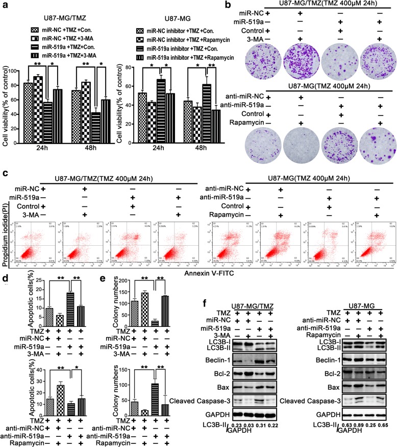
Results: TMZ treatment significantly upregulated miR-519a in U87-MG cells but not in U87-MG/TMZ cells. Moreover, the expression of miR-519a and baseline autophagy levels was lower in U87-MG/TMZ cells as compared to U87-MG cells. miR-519a dramatically enhanced TMZ-induced autophagy and apoptotic cell death in U87-MG/TMZ cells, while inhibition of miR-519a promoted TMZ resistance and reduced TMZ-induced autophagy in U87-MG cells. Furthermore, miR-519a induced autophagy through modification of STAT3 expression. The in vivo results showed that miR-519a can enhance apoptosis and sensitized GBM to TMZ treatment by promoting autophagy and targeting the STAT3/Bcl-2/Beclin-1 pathway. In human GBM tissues, we found an inverse correlation between miR-519a and STAT3 expression.

Conclusions: Our results suggested that miR-519a increased the sensitivity of GBM cells to TMZ therapy. The positive effects of miR-519a may be mediated through autophagy. In addition, miR-519a overexpression can induce autophagy by inhibiting STAT3/Bcl-2 pathway. Therefore, a combination of miR-519a and TMZ may represent an effective therapeutic strategy in GBM.

Keywords: Autophagy; Chemoresistance; Glioblastoma; Signal transducer and activator of transcription 3; miR-519a.


Conflict of interest statement

Ethics approval and consent to participate

This study was approved by the Ethics Committee of Nanfang Hospital. Written informed consent was obtained from all subjects, adhered to the Declaration of Helsinki.

Competing interests

The authors declare that they have no competing interests.

Publisher’s Note

Springer Nature remains neutral with regard to jurisdictional claims in published maps and institutional affiliations.

Figures

Fig. 1
Fig. 1
miR-519a sensitized GBM cells to TMZ treatment. a Parental (black ellipses) or resistant (black squares) cells were exposed to increase the concentrations of TMZ in serum-free medium (acute growth inhibition assay) for 72 h. Cell viability was measured by MTT assays. b The expression of miR-519a was detected by qRT-PCR. c Cell viability of DMSO- or TMZ-treated GBM cells transfected with miR-519a or anti-miR-519a. d, e GBM cells transfected with miR-519a or anti-miR-519a were treated with 400 μM TMZ for 36 h. Cells were harvested and stained with PI and annexin V-FITC for apoptotic analysis (**p < 0.01 vs. TMZ group). f, g Colony formation assay showing the sensitizing effects of miR-519a on GBM cells after TMZ treatment (**p < 0.01 vs. TMZ group). h Western blot analysis of cleaved caspase-3 in GBM cells transfected with miR-519a or anti-miR-519a and then treated with TMZ for 24 h. Data represent the mean (± standard deviations) and are representative of three independent experiments, each performed in triplicate. *p < 0.05, **p < 0.01
Fig. 2
Fig. 2
miR-519a enhanced TMZ-induced autophagy in GBM cells. The expression levels of LC3-II in U87-MG/TMZ and U87-MG cells were evaluated by immunofluorescence assays (a) and Western blotting (b). Red indicates LC3B and blue indicates nuclei. c Both U87-MG/TMZ and U87-MG cells were transfected with GFP-LC3 construct expressing either miR-519a or anti-miR-519a, followed by treatment with 400 μM TMZ. The numbers of GFP-LC3 puncta were quantified using confocal laser scanning microscopy. d Both U87-MG/TMZ and U87-MG cells were transfected with either miR-519a or anti-miR-519a for 24 h, followed by treatment with 400 μM TMZ. Cell samples were prepared for transmission electron microscopy analysis. The arrows indicate autophagic vacuoles. e U87-MG/TMZ cells transfected with miR-519a and parental U87-MG cells transfected with anti-miR-519a were exposed to 400 μM TMZ at different time points (0–24 h). Whole cell lysates were analyzed by Western blotting. f U87-MG/TMZ cells transfected with or without miR-519a and treated with or without 400 μM TMZ after incubation with 20 nM bafilomycin A1 for 2 h. U87-MG cells transfected with or without anti-miR-519a were treated with 20 nM bafilomycin A1 for 2 h. Cell lysates were analyzed by Western blotting. *p < 0.05, **p < 0.01
Fig. 3
Fig. 3
miR-519a sensitized GBM cells to TMZ treatment partly regulated by autophagy. U87-MG/TMZ cells transfected with miR-519a were treated with or without 400 μM TMZ after incubation with 3-MA (an inhibitor of autophagy) for 2 h. U87-MG cells transfected with anti-miR-519a were treated with or without 200 nM rapamycin for 2 h. The cells were then analyzed for the following: a assessment of proliferation by MTT assay; b, e assessment of colony formation; c, d assessment of apoptosis by FACS analysis of PI-stained cells; and f assessment of caspase-3 activity. *p < 0.05, **p < 0.01
Fig. 4
Fig. 4
miR-519a induced autophagy through the modification of STAT3 expression. The expression levels of STAT3 in U87-MG/TMZ and U87-MG cells were evaluated by qRT-PCR (a) and Western blotting (b). U87-MG/TMZ cells were transfected with STAT3 siRNA or negative control siRNA and were subjected to immunoblotting with the indicated antibodies (c). STAT3 knockdown affected GFP-LC3 dot aggregation (d, f). Knockdown of STAT3 affected the number of autophagic vacuoles (e, g). AV (autophagic vacuoles) = autophagosomes and lysosomes. Effects of STAT3 on miR-519a-enhanced autophagy and apoptosis were analyzed by immunoblotting in the indicated cells (h). *p < 0.05, **p < 0.01
Fig. 5
Fig. 5
miR-519a enhanced the antitumor efficacy of TMZ in vivo. The dissected tumors were collected at the end of drug administration (a), and tumor weight was measured (b). The tumor volume was calculated as 4/3 × 3.14 × radius (mm)3 (c). Immunohistochemical analysis (d) and Western blot analysis (e) of phospho-STAT3, LC3B, Bcl-2, Bax, and cleaved caspase-3 levels in xenograft tumors. Xenograft tumors were subjected to TEM. AV (autophagic vacuoles) = autophagosomes and lysosomes (f, g). Coronal T2-weighted MRI of tumors acquired from patient-derived GBM cells (G131212/TMZ) from one animal in each treatment group (red arrow) in the brain samples on day 24 after treatment (h). The survival of mice with orthotopic tumors was measured by Kaplan-Meier survival curves (i). *p < 0.05, **p < 0.01
Fig. 6
Fig. 6
miR-519a was associated with chemoresistance. a miR-519a expression was assessed in primary (n = 24) and recurrent GBM tissue samples (n = 24). b Expression levels of miR-519a were inversely correlated with STAT3 mRNA in tissue samples, as measured by linear regression analysis. c Expression of STAT3, LC3B, and cleaved caspase-3 in primary and recurrent GBM tissue samples were determined by immunochemical staining (magnification × 200). d Schematic illustration of the mechanisms underlying miR-519a induced chemosensitivity to TMZ. Pointed arrows and blunted arrows indicate both activation and repression, respectively. *p < 0.05, **p < 0.01

References

    1. Mostafa H, et al. Immune phenotypes predict survival in patients with glioblastoma multiforme. J Hematol Oncol. 2016;9(1):77. doi: 10.1186/s13045-016-0272-3. - DOI - PMC - PubMed
    1. Stupp R, et al. Radiotherapy plus concomitant and adjuvant temozolomide for glioblastoma. N Engl J Med. 2005;352(10):987–996. doi: 10.1056/NEJMoa043330. - DOI - PubMed
    1. Li B, et al. TMEM140 is associated with the prognosis of glioma by promoting cell viability and invasion. J Hematol Oncol. 2015;8:89. - PMC - PubMed
    1. Wang Z, et al. The D domain of LRRC4 anchors ERK1/2 in the cytoplasm and competitively inhibits MEK/ERK activation in glioma cells. J Hematol Oncol. 2016;9(1):130. doi: 10.1186/s13045-016-0355-1. - DOI - PMC - PubMed
    1. von Ahrens D, et al. The role of stromal cancer-associated fibroblasts in pancreatic cancer. J Hematol Oncol. 2017;10(1):76. doi: 10.1186/s13045-017-0448-5. - DOI - PMC - PubMed

Publication types

MeSH terms

LinkOut - more resources