. 2018 May 29;20(1):95.
doi: 10.1186/s13075-018-1592-1.

Oxidative stress impairs energy metabolism in primary cells and synovial tissue of patients with rheumatoid arthritis

Affiliations

Oxidative stress impairs energy metabolism in primary cells and synovial tissue of patients with rheumatoid arthritis

Emese Balogh et al. Arthritis Res Ther. .

Abstract

Background: In this study, we examined the effect of oxidative stress on cellular energy metabolism and pro-angiogenic/pro-inflammatory mechanisms of primary rheumatoid arthritis synovial fibroblast cells (RASFC) and human umbilical vein endothelial cells (HUVEC).

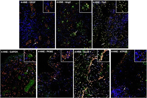
Methods: Primary RASFC and HUVEC were cultured with the oxidative stress inducer 4-hydroxy-2-nonenal (4-HNE), and extracellular acidification rate, oxygen consumption rate, mitochondrial function and pro-angiogenic/pro-inflammatory mechanisms were assessed using the Seahorse analyser, complex I-V activity assays, random mutation mitochondrial capture assays, enzyme-linked immunosorbent assays and functional assays, including angiogenic tube formation, migration and invasion. Expression of angiogenic growth factors in synovial tissue (ST) was assessed by IHC in patients with rheumatoid arthritis (RA) undergoing arthroscopy before and after administration of tumour necrosis factor inhibitors (TNFi).

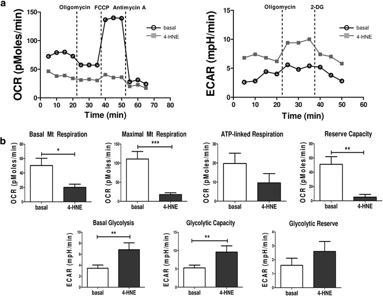
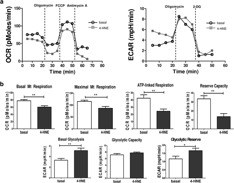
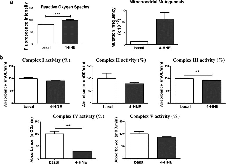
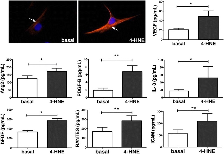
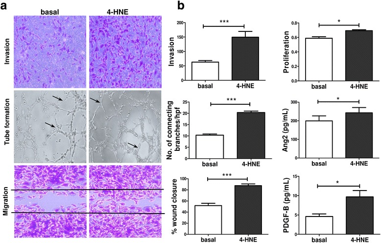
Results: In RASFC and HUVEC, 4-HNE-induced oxidative stress reprogrammed energy metabolism by inhibiting mitochondrial basal, maximal and adenosine triphosphate-linked respiration and reserve capacity, coupled with the reduced enzymatic activity of oxidative phosphorylation complexes III and IV. In contrast, 4-HNE elevated basal glycolysis, glycolytic capacity and glycolytic reserve, paralleled by an increase in mitochondrial DNA mutations and reactive oxygen species. 4-HNE activated pro-angiogenic responses of RASFC, which subsequently altered HUVEC invasion and migration, angiogenic tube formation and the release of pro-angiogenic mediators. In vivo markers of angiogenesis (vascular endothelial growth factor, angiopoietin 2 [Ang2], tyrosine kinase receptor [Tie2]) were significantly associated with oxidative damage and oxygen metabolism in the inflamed synovium. Significant reduction in ST vascularity and Ang2/Tie2 expression was demonstrated in patients with RA before and after administration of TNFi.

Conclusions: Oxidative stress promotes metabolism in favour of glycolysis, an effect that may contribute to acceleration of inflammatory mechanisms and subsequent dysfunctional angiogenesis in RA.

Keywords: Angiogenesis; Bioenergetic metabolism; Oxidative stress; Rheumatoid arthritis.


Conflict of interest statement

Ethics approval and consent to participate

All of this research was carried out in accordance with the Declaration of Helsinki, and approval for this study was granted by the St. Vincent’s University Hospital Medical Research and Ethics Committee. All patients gave fully informed written consent approved by the institutional ethics committee.

Consent for publication

Written consent was obtained from all the participants in and authors of this study.

Competing interests

The authors declare that they have no competing interests.

Publisher’s Note

Springer Nature remains neutral with regard to jurisdictional claims in published maps and institutional affiliations.

Figures

Fig. 1
Fig. 1
Bioenergetic metabolism in primary rheumatoid arthritis synovial fibroblast cells (RASFC) subjected to 4-hydroxy-2-nonenal (4-HNE)-induced oxidative stress. a Representative oxygen consumption rate (OCR) and extracellular acidification rate (ECAR) Seahorse analyser profiles before and after injections of oligomycin, trifluorocarbonylcyanide phenylhydrazone (FCCP), antimycin A and 2-deoxyglucose (2-DG) in RASFC in the presence and absence of 4-HNE. b Bar graphs demonstrate quantification of basal mitochondrial (Mt) respiration, maximal Mt respiration, adenosine triphosphate (ATP) synthesis, reserve capacity, basal glycolysis, glycolytic capacity and glycolytic reserve in RASFC (n = 5) subjected to oxidative stress. Data are presented as mean ± SEM. *p < 0.05, **p < 0.01, and ***p < 0.001, significant differences from basal level
Fig. 2
Fig. 2
Bioenergetic metabolism in human umbilical vein endothelial cells (HUVEC) subjected to 4-hydroxy-2-nonenal (4-HNE)-induced oxidative stress. a Representative oxygen consumption rate (OCR) and extracellular acidification rate (ECAR) Seahorse analyser profiles before and after injections of oligomycin, trifluorocarbonylcyanide phenylhydrazone (FCCP), antimycin A and 2-deoxyglucose (2-DG) in HUVEC in the presence and absence of 4-HNE. b Bar graphs demonstrate quantification of basal mitochondrial (Mt) respiration, maximal Mt respiration, adenosine triphosphate (ATP) synthesis, reserve capacity, basal glycolysis, glycolytic capacity and glycolytic reserve in HUVEC (n = 3) subjected to oxidative stress. Data are presented as mean ± SEM. *p < 0.05 and **p < 0.01, significant differences from basal level
Fig. 3
Fig. 3
Mitochondrial mutagenesis and activity of enzymes of mitochondrial oxidative phosphorylation (OXPHOS) complexes under 4-hydroxy-2-nonenal (4-HNE)-induced oxidative stress. a Bar graphs demonstrate increased production of reactive oxygen species (n = 7), paralleled by the greater frequency of mitochondrial DNA mutation (n = 5) in primary rheumatoid arthritis synovial fibroblast cells (RASFC) in response to 4-HNE. b Activity of mitochondrial OXPHOS complexes I–V in the presence of 4-HNE. 4-HNE reduces the activity of complex I by 9%, complex II by 22%, complex III by 8%, complex IV by 70% and complex V by 12% (all complexes measured in triplicate). For each complex, results are graphically demonstrated as the percentage of enzymatic activity in the presence of 4-HNE relative to the percentage of basal activity. Data is represented as Mean ± SEM, **p<0.01; ***p<0.001 significantly different to basal
Fig. 4
Fig. 4
4-Hydroxy-2-nonenal (4-HNE) induces pro-angiogenic and pro-inflammatory mechanisms in primary rheumatoid arthritis synovial fibroblast cells (RASFC). Increased vascular endothelial growth factor (VEGF) immunofluorescence in RASFC subjected to 4-HNE compared to the basal cells and quantification of VEGF, angiopoietin 2 (Ang2), basic fibroblast growth factor (bFGF), interleukin (IL)-8, platelet-derived growth factor subunit B (PDGF-B), regulated on activation, normal T cell expressed and secreted (RANTES), intercellular adhesion molecule (ICAM) in RASFC supernatants (n = 7) following cell culture with 4-HNE. Data are presented as mean ± SEM. *p < 0.05 and **p < 0.01, significant differences from basal level. Red = VEGF; blue = 4′,6-diamidino-2-phenylindole–; magnification of photomicrographs × 40
Fig. 5
Fig. 5
Effects of primary rheumatoid arthritis synovial fibroblast cell (RASFC)-conditioned media on angiogenic responses of human umbilical vein endothelial cells (HUVEC). a Representative images demonstrating invasion, the formation of tube-like structures and migration of HUVEC cultured in the presence of basal or 4-hydroxy-2-nonenal (4-HNE)-supplemented conditioned media. Magnification × 10 of photomicrographs demonstrating invasion, tube formation (arrows indicate connecting branches) and cell migration. b Bar graphs demonstrate an increase in the number of invading, proliferating and migrating HUVEC, a higher number of connecting branches formed between HUVEC, and greater angiopoietin 2 (Ang2) and platelet-derived growth factor subunit B (PDGF-B) release from HUVEC exposed to 4-HNE-supplemented conditioned media (n = 6). Data are presented as mean ± SEM. *p < 0.05 and ***p < 0.001, significant differences from basal level. hpf High-power field
Fig. 6
Fig. 6
Synovial tissue (ST) angiogenesis, oxidative stress and cellular bioenergetics. To support the concept that oxidative stress, angiogenesis and energy metabolism are interconnected processes that co-exist during the inflammation milieu, double-immunofluorescence staining was performed. ST slides were co-incubated with primary mouse antibody against human 4-hydroxy-2-nonenal (4-HNE) and with primary rabbit antibodies against angiogenic factors (vascular endothelial growth factor [VEGF], angiopoietin 2 [Ang2], tyrosine kinase receptor [Tie2]), glycolytic proteins (glyceraldehyde 3-phosphate dehydrogenase [GAPDH], pyruvate kinase isozyme 2 [PKM2], glucose transporter 1 [GLUT1]) and a mitochondrial marker (adenosine triphosphate synthase subunit β [ATP5B]). Representative merged immunofluorescence images demonstrate examples of co-localisation (yellow) of 4-HNE with VEGF, Ang2, Tie2, GAPDH, PKM2, GLUT1 and ATP5B. Cells stained green are positive for 4-HNE only; cells stained red are positive only for VEGF, Ang2, Tie2, GAPDH, PKM2, GLUT1 and ATP5B. Arrows indicate examples of co-localisation. Magnification of photomicrographs × 20, insets show high-power magnification of co-localisation. Representative images show single immunofluorescence of 4-HNE, VEGF, Ang2, Tie2, GAPDH, PKM2, GLUT1 and ATP5B along with their controls. Isotype-matched antibodies are shown in Additional file 3: Figure S3 and Additional file 4: Figure S4

References

    1. Tas SW, Maracle CX, Balogh E, Szekanecz Z. Targeting of proangiogenic signalling pathways in chronic inflammation. Nat Rev Rheumatol. 2016;12(2):111–122. doi: 10.1038/nrrheum.2015.164. - DOI - PubMed
    1. Bodamyali T, Stevens CR, Billingham ME, Ohta S, Blake DR. Influence of hypoxia in inflammatory synovitis. Ann Rheum Dis. 1998;57(12):703–710. doi: 10.1136/ard.57.12.703. - DOI - PMC - PubMed
    1. Stevens CR, Blake DR, Merry P, Revell PA, Levick JR. A comparative study by morphometry of the microvasculature in normal and rheumatoid synovium. Arthritis Rheum. 1991;34(12):1508–1513. doi: 10.1002/art.1780341206. - DOI - PubMed
    1. Taylor RW, Turnbull DM. Mitochondrial DNA mutations in human disease. Nat Rev Genet. 2005;6(5):389–402. doi: 10.1038/nrg1606. - DOI - PMC - PubMed
    1. Ospelt C, Gay S. Somatic mutations in mitochondria: the chicken or the egg? Arthritis Res Ther. 2005;7(5):179–180. doi: 10.1186/ar1809. - DOI - PMC - PubMed

Publication types

MeSH terms