. 2018 Jul 20;293(29):11283-11295.
doi: 10.1074/jbc.RA118.002182. Epub 2018 May 29.

An allosteric propofol-binding site in kinesin disrupts kinesin-mediated processive movement on microtubules

Affiliations

An allosteric propofol-binding site in kinesin disrupts kinesin-mediated processive movement on microtubules

Kellie A Woll et al. J Biol Chem. .

Abstract

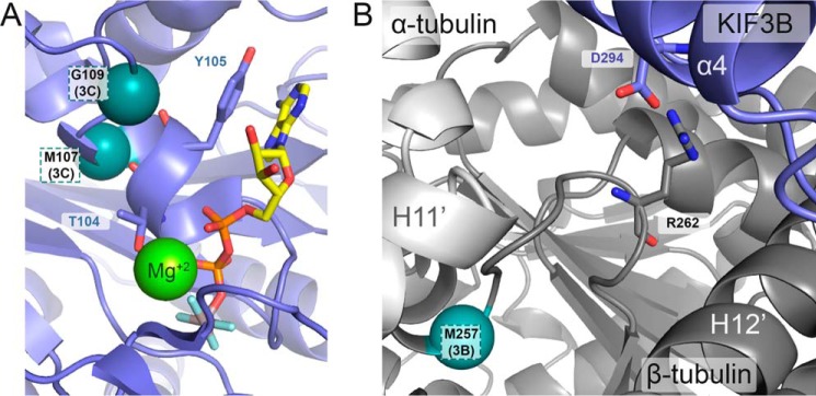
Microtubule-based molecular motors mediate transport of intracellular cargo to subdomains in neurons. Previous evidence has suggested that the anesthetic propofol decreases the average run-length potential of the major anterograde transporters kinesin-1 and kinesin-2 without altering their velocity. This effect on kinesin has not been observed with other inhibitors, stimulating considerable interest in the underlying mechanism. Here, we used a photoactive derivative of propofol, meta-azipropofol (AziPm), to search for potential propofol-binding sites in kinesin. Single-molecule motility assays confirmed that AziPm and propofol similarly inhibit kinesin-1 and kinesin-2. We then applied AziPm in semiquantitative radiolabeling and MS microsequencing assays to identify propofol-binding sites within microtubule-kinesin complexes. The radiolabeling experiments suggested preferential AziPm binding to the ATP-bound microtubule-kinesin complex. The photolabeled residues were contained within the kinesin motor domain rather than at the motor domain-β-tubulin interface. No residues within the P-loop of kinesin were photolabeled, indicating an inhibitory mechanism that does not directly affect ATPase activity and has an effect on run length without changing velocity. Our results also indicated that when the kinesin motor interacts with the microtubule during its processive run, a site forms in kinesin to which propofol can then bind and allosterically disrupt the kinesin-microtubule interaction, resulting in kinesin detachment and run termination. The discovery of the propofol-binding allosteric site in kinesin may improve our understanding of the strict coordination of the motor heads during the processive run. We hypothesize that propofol's potent effect on intracellular transport contributes to various components of its anesthetic action.

Keywords: anesthesia; kinesin; microtubule; molecular motor; photoaffinity labeling; propofol; single molecule motility; single-molecule biophysics; tubulin.


Conflict of interest statement

The authors declare that they have no conflicts of interest with the contents of this article

Figures

Figure 1.
Figure 1.
Generalized schematic of the kinesin stepping cycle with proposed states for propofol-induced premature detachment from the microtubule. E0, dimeric kinesin in solution, detached from the microtubule holds ADP tightly bound in each motor head. E0–E1, the processive run starts with microtubule collision followed by ADP release. The leading head is in the no-nucleotide state (white; Ø) and the trailing head is detached from the microtubule with ADP tightly bound. E2–E4, ATP binding at the leading head initiates a series of structural transitions, namely the ridged movement of the P-loop and Switch I/II motor head subdomains and neck linker docking that promotes the trailing head to move forward to the next microtubule-binding site. E4–E5, ADP is released from the leading head resulting in a two-head bound state. Strain develops, the leading head neck linker is detached and pointed backward, which decreases the probability of ATP binding to the leading nucleotide-free head. E5–E1, ATP hydrolysis within the trailing head followed Pi release weakens the affinity of the trailing head to the microtubule, resulting in its subsequent detachment. The leading head is now able to bind another ATP to continue the processive run. The states most vulnerable to propofol-induced detachment from the microtubule include E1–E4 based on the AziPm photolabeling.
Figure 2.
Figure 2.
AziPm-like propofol disrupts kinesin-1 processivity without an impact on velocity. A, comparison of the chemical structure of propofol with its photoaffinity derivative, AziPm. B–D, single molecule K439 run length and velocity (inset) data and representative kymographs of Qdot-labeled K439 motors in (B) 5% DMSO control, (C) 10 ìM propofol, and (D) 10 μm AziPm. A single exponential decay fit provides the mean run length ± S.E. for each dataset. Mean run-length differences between the DMSO control and either propofol or AziPm were highly significant (p ≪ 0.0001), yet the mean run lengths of propofol and AziPm conditions showed no statistical significance from each other (p > 0.3). A Gaussian fit provides the mean velocity ± S.E. for each dataset, which were not statistically significant between the DMSO control and either propofol or AziPm (p > 0.1). Kymograph scale bars: 5 μm along the x axis, 25 s along the y axis.
Figure 3.
Figure 3.
Photolabeled residues in kinesin-1 and kinesin-2 motor domains. Shown is alignment of kinesin motor domain sequences from kinesin-1 (K439) and kinesin-2 (KIF3B and KIF3C), with structural elements from kinesin-1 (KIF5B, PDB code 4HNA) provided above the alignment (30). Residues highlighted in magenta were photolabeled by AziPm in the no-nucleotide state for K439 (Phe318) and KIF3C (Leu242, Asp330, Leu332). Residues in the ATP-like AMPPNP-bound state within the common allosteric binding site are highlighted in cyan for K439 (C7, Leu227, Ser289), KIF3B (Tyr138), and KIF3C (Ser144, Leu242, Asn243). The KIF3C residue Leu242 was photolabeled by AziPm in the absence of nucleotide and in the presence of 1 mm AMPPNP, the ATP-like state (cyan with magenta underline). Additionally, residues photolabeled at a site unique to KIF3C in the presence of AMPPNP (Met107, Gly109) are highlighted in teal.
Figure 4.
Figure 4.
Location of predicted propofol and AziPm-binding pockets and associated photolabeled residues in microtubule–kinesin complexes without and with AMPPNP. A, side and focused view of the X-ray crystal structure of kinesin motor head in complex with tubulin in the absence of nucleotide (PDB ID 4LNU) (29) respresenting photolabeled microtubule–kinesin complexes without (−) AMPPNP. The green Connolly surface representations highlight the highest scored poses for five propofol and five AziPm as predicted by AutoDock Vina (46) within the CASTp (45) predicted binding cavity (see appendix Fig. S11). Magenta spheres indicate the α-carbon atoms of Phe318 photolabeled in K439 and Asp288 and Leu290 that correspond with the photolabeled residues Asp330 and Leu332 in KIF3C, respectively. B, side and focused view of the X-ray crystal structure of ADP-AlF4-bound kinesin motor head in complex with tubulin (PDB ID 4HNA) (30) representing photolabeled microtubule–kinesin complexes with (+) AMPPNP. The orange Connolly surface representations highlight five propofol and five AziPm in the highest scored poses predicted by AutoDock Vina (46) within the CASTp (45) predicited binding cavity (see Fig. S14). Blue spheres indicate the α-carbon atoms of the photolabeled residue in K439 (Ser289), photolabeled residues in KIF3C (Ser144 and Asn243) corresponding with Ser133 and Tyr226 in K439, respectively, and the photolabeled residue in both K439 (Leu227) and KIF3C (corresponding with Leu242). ADP-AlF4 is shown in stick representation and a Mg2+ ion is shown as a light green sphere. All residues within predicted binding sites were made flexible in AutoDock Vina, whereas the backbone structure remained rigid during docking experiments.
Figure 5.
Figure 5.
AziPm photolabeled residue identified in secondary sites for KIF3C and KIF3AB β-tubulin within the AMPPNP-bound microtubule–kinesin complexes. A and B, expanded views of the X-ray crystal structure of ADP-AlF4-bound kinesin motor head in complex with tubulin (PDB ID 4HNA) to represent the microtubule–kinesin complexes with AMPPNP. A, photolabeled residues (Met107 and Gly109) within the KIF3C second the photolabeled pocket from the nucleotide-binding site are labeled accordingly. ADP-AlF4 is shown in stick representation and a magnesium ion is shown as a light green sphere. B, photoaffinity labeled residue (Met257) in β-tubulin within KIF3AB microtubule–kinesin complexes with AMPPNP and the residue's relationship to TUBB3 Arg262. The α-carbon of photolabeled residues are represented as teal spheres and are labeled accordingly.

References

    1. Bentley M., and Banker G. (2016) The cellular mechanisms that maintain neuronal polarity. Nat. Rev. Neurosci. 17, 611–622 10.1038/nrn.2016.100 - DOI - PubMed
    1. Hirokawa N., Niwa S., and Tanaka Y. (2010) Molecular motors in neurons: transport mechanisms and roles in brain function, development, and disease. Neuron 68, 610–638 10.1016/j.neuron.2010.09.039 - DOI - PubMed
    1. Maday S., Twelvetrees A. E., Moughamian A. J., and Holzbaur E. L. (2014) Axonal transport: cargo-specific mechanisms of motility and regulation. Neuron 84, 292–309 10.1016/j.neuron.2014.10.019 - DOI - PMC - PubMed
    1. Hirokawa N., Noda Y., Tanaka Y., and Niwa S. (2009) Kinesin superfamily motor proteins and intracellular transport. Nat. Rev. Mol. Cell Biol. 10, 682–696 10.1038/nrm2774 - DOI - PubMed
    1. Miki H., Setou M., Kaneshiro K., and Hirokawa N. (2001) All kinesin superfamily protein, KIF, genes in mouse and human. Proc. Natl. Acad. Sci. U.S.A. 98, 7004–7011 10.1073/pnas.111145398 - DOI - PMC - PubMed

Publication types

LinkOut - more resources