Review
. 2018 Nov 1;10(11):a032797.
doi: 10.1101/cshperspect.a032797.

Riboswitches and Translation Control

Affiliations
Review

Riboswitches and Translation Control

Ronald R Breaker. Cold Spring Harb Perspect Biol. .

Abstract

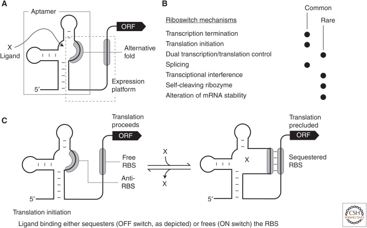
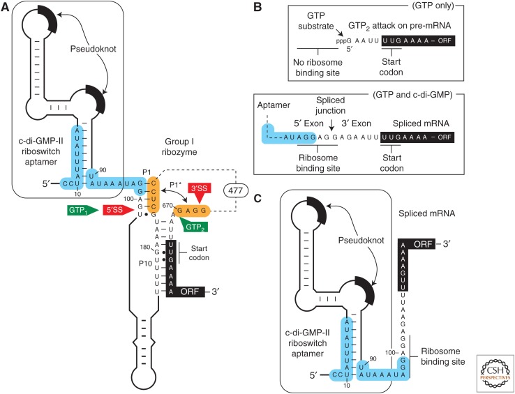
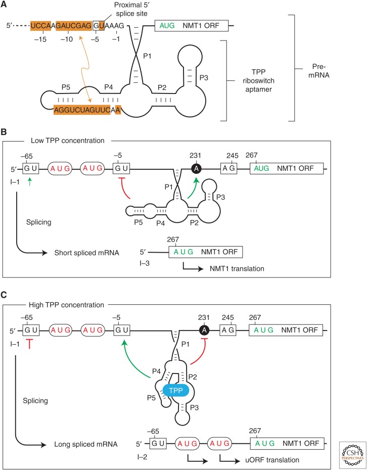
A growing collection of bacterial riboswitch classes is being discovered that sense central metabolites, coenzymes, and signaling molecules. Included among the various mechanisms of gene regulation exploited by these RNA regulatory elements are several that modulate messenger RNA (mRNA) translation. In this review, the mechanisms of riboswitch-mediated translation control are summarized to highlight both their diversity and potential ancient origins. These mechanisms include ligand-gated presentation or occlusion of ribosome-binding sites, control of alternative splicing of mRNAs, and the regulation of mRNA stability. Moreover, speculation on the potential for novel riboswitch discoveries is presented, including a discussion on the potential for the discovery of a greater diversity of mechanisms for translation control.


Figures

Figure 1.
Figure 1.
Schematic representations of common riboswitch expression platform arrangements. (A) Riboswitches typically carry a single ligand-binding aptamer (gray box) located upstream of (and slightly overlapping) the expression platform (dashed box). Folding changes in the aptamer, brought about by ligand binding, cause folding changes in the expression platform to regulate gene expression by various mechanisms. (B) List of experimentally validated or predicted riboswitch gene-control mechanisms. Processes by which the mechanisms highlighted in bold italic font regulate translation and are discussed in the text. (C) Schematic representation of a riboswitch that permits translation in the absence of ligand (left), but inhibits translation when bound to ligand (right). In the model depicted, ligand binding sequesters the ribosome-binding site (RBS) and prevents ribosome binding to the messenger RNA (mRNA). Alternatively, some riboswitch RNAs liberate the RBS on ligand binding to promote ribosome binding and translation.
Figure 2.
Figure 2.
Regulation of self-splicing ribozyme function by a riboswitch aptamer. (A) Schematic representation of the 5′ untranslated region (5′UTR) of the CD3246 gene from Clostridium difficile, which carries a c-di-GMP-II riboswitch aptamer immediately upstream of a group I self-splicing ribozyme. The binding of c-di-GMP to the aptamer domain requires the structure depicted. This structure promotes ribozyme-catalyzed attack of GTP (called GTP1) at the 5′ splice site (5′SS) and subsequent attack of the 5′ exon’s 3′ terminus oxygen on the phosphorus of the 3′ splice site (3′SS) to yield a properly spliced messenger RNA (mRNA) product that can be translated. In the absence of c-di-GMP binding, portions of the aptamer and ribozyme shaded in blue reorganize to form an alternative base-paired structure. This disrupts the P1 stem that is required for proper splicing, and permits the formation of P1* to present an alternative site for GTP attack (called GTP2). GTP attack at this alternative site removes all but four nucleotides located upstream of the unusual UUG start codon for the ORF. (B) Two reaction products produced by the tandem aptamer-ribozyme construct depicted in A. (Top) Processed mRNA product generated by ribozyme action in the absence of c-di-GMP binding. Note that the ribosome-binding site (RBS) has been removed, and thus translation of the downstream open reading frame (ORF) is precluded. (Bottom) Processed mRNA product resulting from ribozyme splicing in the presence of c-di-GMP. Splicing creates a strong RBS that permits translation to occur. (C) Although the properly spliced mRNA (B, bottom) carries an RBS, the riboswitch aptamer presumably can still operate to occlude or reveal this sequence based on the availability of c-di-GMP. Specifically, the nucleotides shaded in blue that alternatively pair to regulate ribozyme splicing activity remain present in the properly spliced mRNA, and presumably can be exploited as an expression platform to continue to regulate gene expression as would a more common riboswitch.
Figure 3.
Figure 3.
Translation regulation by a fungal thiamin pyrophosphate (TPP) riboswitch that controls alternative splicing. (A) Schematic representation of the region of the Neurospora crassa NMT1 pre–messenger RNA (mRNA) region that includes an intron and TPP riboswitch. The TPP aptamer (including stems P1 through P5) is stabilized on the binding ligand as depicted, or nucleotides from the P4 and P5 region can alternatively base-pair to a region encompassing a 5′SS. (B) Results of mRNA splicing when the TPP concentration is low. The alternative base-pairing depicted in A will sequester the proximal 5′SS to promote the use of a distal 5′SS. This produces a short mRNA splicing product wherein the main NMT1 open reading frame (ORF) can be translated. (C) TPP binding to the aptamer prevents occlusion of the proximal 5′SS, which results in a longer mRNA splicing product. This alternatively spliced RNA carries upstream ORFs (uORFs) that are translated instead of the main NMT1 ORF.

References

    1. Ames TD, Breaker RR. 2010. Bacterial riboswitch diversity and analysis. In The chemical biology of nucleic acids (ed. Mayer G), Chap. 20 Wiley, Chichester, UK.
    1. Antson AA, Dodson EJ, Dodson G, Greaves RB, Chen X, Gollnick P. 1999. Structure of the trp RNA-binding attenuation protein, TRAP, bound to RNA. Nature 401: 235–242. - PubMed
    1. Arachchilage GM, Sherlock ME, Weinberg Z, Breaker RR. 2017. SAM-VI RNAs selectively bind S-adenosylmethionine and exhibit similarities to SAM-III riboswitches. RNA Biol 10.1080/15476286.2017.1399232. - DOI - PMC - PubMed
    1. Barrick JE, Breaker RR. 2007. The distributions, mechanisms, and structures of metabolite-binding riboswitches. Genome Biol 8: R239. - PMC - PubMed
    1. Barrick JE, Corbino KA, Winkler WC, Nahvi A, Mandal M, Collins J, Lee M, Roth A, Sudarsan N, Jona I, et al. 2004. New RNA motifs suggest an expanded scope for riboswitches in bacterial genetic control. Proc Natl Acad Sci 101: 6421–6426. - PMC - PubMed

Publication types