. 2018 Jun 26;115(26):6810-6815.
doi: 10.1073/pnas.1718811115. Epub 2018 May 29.

Diverse AR-V7 cistromes in castration-resistant prostate cancer are governed by HoxB13

Affiliations

Diverse AR-V7 cistromes in castration-resistant prostate cancer are governed by HoxB13

Zhong Chen et al. Proc Natl Acad Sci U S A. .

Erratum in

Abstract

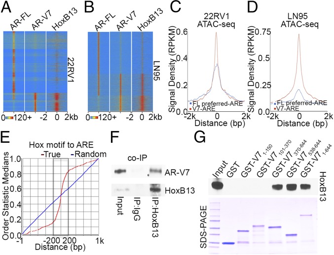
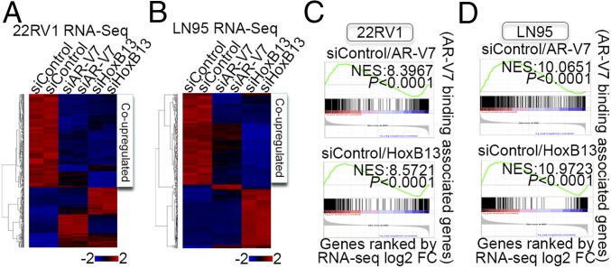
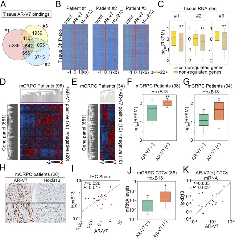
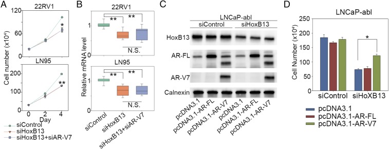
The constitutively active androgen receptor (AR) splice variant 7 (AR-V7) plays an important role in the progression of castration-resistant prostate cancer (CRPC). Although biomarker studies established the role of AR-V7 in resistance to AR-targeting therapies, how AR-V7 mediates genomic functions in CRPC remains largely unknown. Using a ChIP-exo approach, we show AR-V7 binds to distinct genomic regions and recognizes a full-length androgen-responsive element in CRPC cells and patient tissues. Remarkably, we find dramatic differences in AR-V7 cistromes across diverse CRPC cells and patient tissues, regulating different target gene sets involved in CRPC progression. Surprisingly, we discover that HoxB13 is universally required for and colocalizes with AR-V7 binding to open chromatin across CRPC genomes. HoxB13 pioneers AR-V7 binding through direct physical interaction, and collaborates with AR-V7 to up-regulate target oncogenes. Transcriptional coregulation by HoxB13 and AR-V7 was further supported by their coexpression in tumors and circulating tumor cells from CRPC patients. Importantly, HoxB13 silencing significantly decreases CRPC growth through inhibition of AR-V7 oncogenic function. These results identify HoxB13 as a pivotal upstream regulator of AR-V7-driven transcriptomes that are often cell context-dependent in CRPC, suggesting that HoxB13 may serve as a therapeutic target for AR-V7-driven prostate tumors.

Keywords: AR-V7; HoxB13; castration-resistant prostate cancer; motif-resolution cistromes.


Conflict of interest statement

The authors declare no conflict of interest.

Figures

Fig. 1.
Fig. 1.
AR-V7 cistromes and their regulated transcriptomes are heterogeneous in AR-V7–driven CRPC. (A) A heatmap of differentially expressed genes [false-discovery rate (FDR) < 0.05, fold-change > 2] after AR-V7 silencing. The gene expression [reads per kilobase per million mapped reads (RPKM)] values for each gene were normalized to the standard normal distribution to generate z-scores. The scale bar is shown with the minimum expression value for each gene in blue and the maximum value in red. (B) Venn diagrams show AR-V7 up-regulated and down-regulated genes in 22RV1 and LN95 cells, respectively. (C) Overlap of AR-V7 binding sites between 22RV1 and LN95 cells. (D and E) ChIP-exo raw tags distribution (1-bp resolution) over AREs on the forward (blue, Left) and reverse (red, Right) strands, respectively in 22RV1 cells (D) and LN95 cells (E). The Center panels represent the bound ARE sequences ordered as in the Left and Right.
Fig. 2.
Fig. 2.
AR-V7 and HoxB13 interact genomically and physically. (A and B) Heatmaps show the ChIP-exo signal intensity of AR-FL, AR-V7, and HoxB13 binding in 22RV1 cells (A) and LN95 cells (B). (C and D) Chromatin accessibility in 22RV1 (C) and LN95 (D) cells was determined by ATAC-seq. Normalized averaged ATAC-seq signal tag distribution over AR-V7 ARE regions and AR-FL preferred-ARE regions is shown. The window indicates ±2-kb regions of AREs. (E) Cumulative genomic position distributions of precisely defined HoxB13 motifs relative to AR-V7–bound AREs in AR-V7/HoxB13 binding locations in 22RV1 and LN95 cells. (F) Whole-cell lysates from hormone-depleted 22RV1 cells were immunoprecipitated with HoxB13 antibodies, and Western blots were performed with antibodies against AR-V7 and HoxB13. (G) GST pull-down assays were performed by incubating in vitro translated HoxB13 with GST ∼ AR-V7 fusion proteins. Western blots were performed using an anti-HoxB13 antibody.
Fig. 3.
Fig. 3.
HoxB13 and AR-V7 coup-regulate diverse target oncogenes in CRPC. (A and B) Heatmaps of regulated genes (FDR < 0.05, fold-change > 2) after AR-V7 or HoxB13 silencing in 22RV1 (A) and LN95 cells (B). (C and D) GSEA analyses compare associated genes within ±50 kb of AR-V7 binding locations with AR-V7 or HoxB13 regulated genes determined by RNA-seq analysis in 22RV1 (C) and LN95 (D) cells.
Fig. 4.
Fig. 4.
HoxB13 and AR-V7 coregulate transcriptomic diversity among CRPC patients. (A) A Venn diagram shows AR-V7 binding site diversity between the three CRPC patients. (B) Heatmaps show the ChIP-exo signal intensity of AR-V7 and HoxB13 binding in three CRPC patients. (C) Box plots compare expression of genes associated with tissue AR-V7/HoxB13 binding sites. Coup-regulated genes refer to genes that were also coup-regulated by AR-V7/HoxB13 in CRPC cells. (D and E) Heatmaps show expression of the 681-gene panel among the 98 metastatic CRPC (mCRPC) patients in the Robinson et al. (20) cohort (D), and 34 mCRPC cases in the Beltran et al. (21) cohort (E). (F and G) Box plots compare HoxB13 gene expression between AR-V7+ and AR-V7 patients in the Robinson et al. (20) cohort (F) and in the Beltran et al. (21) cohort (G), respectively. The significance was determined by Mann–Whitney rank sum test. *P < 0.01, **P < 0.001. (H) Representative AR-V7 and HoxB13 immunoreactivity in mCRPC tissues (Upper 20×, Lower 40× original magnification). (I) Correlation between AR-V7 and HoxB13 staining in 20 CRPC cases. Slides were scanned using an Aperio Digital Pathology Slide Scanner (Leica Biosystems) at 40× magnification and staining quantified using the Aperio Image Scope (v11). Data represent the ratio of positive pixels per total pixels from areas of tissue containing mCRPC. (J) A box plot compares HoxB13 mRNA levels in 86 CTCs from mCRPC patients. The significance was determined by one-tailed t test. *P < 0.05. (K) Correlation of mRNA expression between HoxB13 and AR-V7 in AR-V7+ CTCs.
Fig. 5.
Fig. 5.
AR-V7 is an important mediator of HoxB13 function in CRPC. (A and B) 22RV1 cells and LN95 cells were transfected with control siRNAs, siRNAs targeting HoxB13, and combined siRNAs targeting HoxB13 and AR-V7. Cell proliferation was measured by direct cell count assays (A), and expression of HoxB13 and AR-V7 coregulated genes AAK1, CROT, NEDD4L, and GRIN3A was analyzed by RT-PCR (B). The results for cell proliferation (A) are shown as mean ± SD (n = 2). The significance for cell proliferation (A) and RT-PCR (B) was determined by one-way ANOVA. *P < 0.01, **P < 0.001. (C and D) LNCaP-abl cells were cotransfected with siRNAs combined with pcDNA3.1 vector, pcDNA3.1–AR-FL (32), or pcDNA3.1–AR-V7 (11). Western blots were performed using the indicated antibodies (C), and cell proliferation on day 5 was measured by direct cell count assays (D). The significance was determined by one-way ANOVA. *P < 0.01.
Fig. 6.
Fig. 6.
HoxB13 silencing inhibits AR-V7-driven CRPC growth in vivo. (A and D) HoxB13 protein expression in 22RV1 cells (A) and LN95 cells (D) infected with lentivirus encoding HoxB13 shRNA or a control shRNA after 48 h. Western blots were performed using the same cells used for injection into castrated mice. (B and E) Representative picture of shControl 22RV1 tumors and HoxB13-silenced 22RV1 tumors at time of collection (B, day 24), and shControl LN95 tumors and HoxB13-silenced LN95 tumors at time of collection (E, day 34). (C and F) Average 22RV1 tumor weight for control (C, n = 24) and HoxB13 silenced groups (C, n = 23), and average LN95 tumor weight for control (F, n = 24) and HoxB13 silenced groups (F, n = 22). The results are shown as mean ± SE. The significance was determined by one-tailed t test. *P < 0.01, **P < 0.001. (G and H) AR-V7 and HoxB13 tissue ChIP analysis using engrafted 22RV1 (G, tumors n = 12, regions n = 8) and LN95 (H, tumors n = 20, regions n = 8) tumor tissues. The significance was determined by Mann–Whitney rank sum test. **P < 0.001.

Comment in

References

    1. Feldman BJ, Feldman D. The development of androgen-independent prostate cancer. Nat Rev Cancer. 2001;1:34–45. - PubMed
    1. Watson PA, Arora VK, Sawyers CL. Emerging mechanisms of resistance to androgen receptor inhibitors in prostate cancer. Nat Rev Cancer. 2015;15:701–711. - PMC - PubMed
    1. Mangelsdorf DJ, et al. The nuclear receptor superfamily: The second decade. Cell. 1995;83:835–839. - PMC - PubMed
    1. Maughan BL, Antonarakis ES. Clinical relevance of androgen receptor splice variants in castration-resistant prostate cancer. Curr Treat Options Oncol. 2015;16:57. - PubMed
    1. Antonarakis ES, Armstrong AJ, Dehm SM, Luo J. Androgen receptor variant-driven prostate cancer: Clinical implications and therapeutic targeting. Prostate Cancer Prostatic Dis. 2016;19:231–241. - PMC - PubMed

Publication types

MeSH terms