. 2018 Jul;29(7):1875-1886.
doi: 10.1681/ASN.2017080828. Epub 2018 May 29.

Mechanisms of Crystalloid versus Colloid Osmosis across the Peritoneal Membrane

Affiliations

Mechanisms of Crystalloid versus Colloid Osmosis across the Peritoneal Membrane

Johann Morelle et al. J Am Soc Nephrol. 2018 Jul.

Abstract

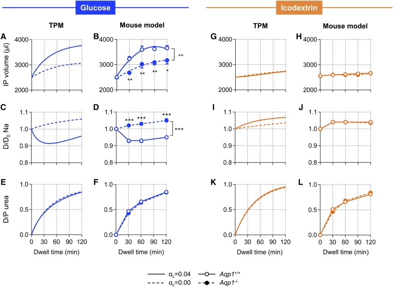
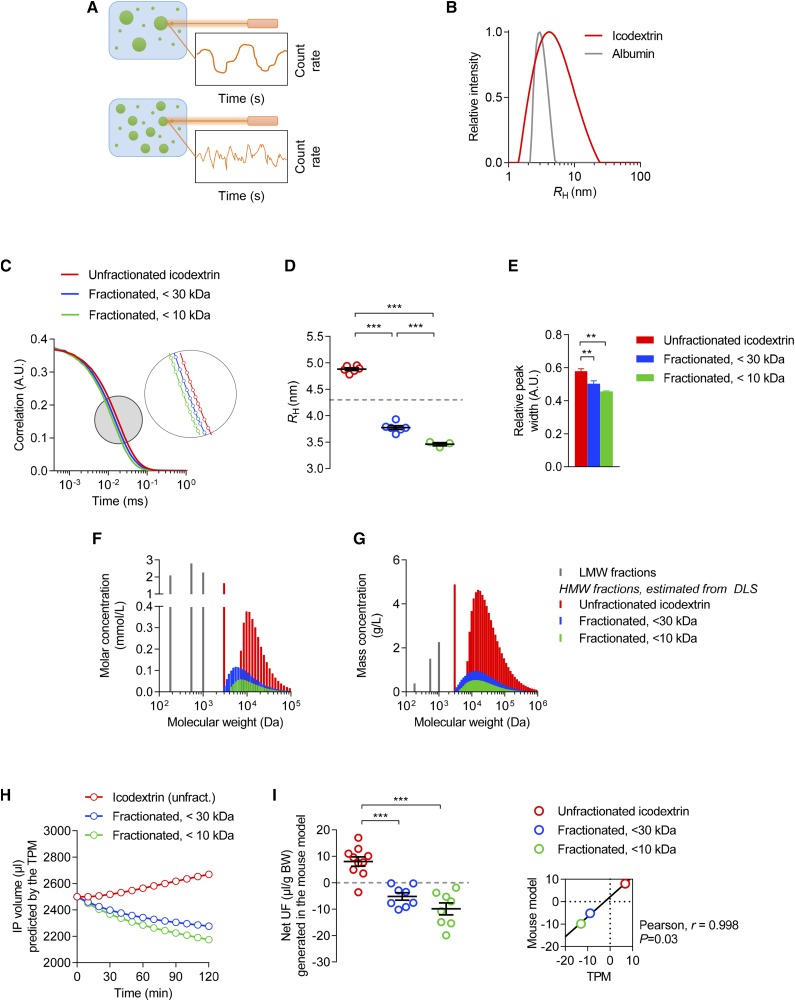
Background Osmosis drives transcapillary ultrafiltration and water removal in patients treated with peritoneal dialysis. Crystalloid osmosis, typically induced by glucose, relies on dialysate tonicity and occurs through endothelial aquaporin-1 water channels and interendothelial clefts. In contrast, the mechanisms mediating water flow driven by colloidal agents, such as icodextrin, and combinations of osmotic agents have not been evaluated.Methods We used experimental models of peritoneal dialysis in mouse and biophysical studies combined with mathematical modeling to evaluate the mechanisms of colloid versus crystalloid osmosis across the peritoneal membrane and to investigate the pathways mediating water flow generated by the glucose polymer icodextrin.ResultsIn silico modeling and in vivo studies showed that deletion of aquaporin-1 did not influence osmotic water transport induced by icodextrin but did affect that induced by crystalloid agents. Water flow induced by icodextrin was dependent upon the presence of large, colloidal fractions, with a reflection coefficient close to unity, a low diffusion capacity, and a minimal effect on dialysate osmolality. Combining crystalloid and colloid osmotic agents in the same dialysis solution strikingly enhanced water and sodium transport across the peritoneal membrane, improving ultrafiltration efficiency over that obtained with either type of agent alone.Conclusions These data cast light on the molecular mechanisms involved in colloid versus crystalloid osmosis and characterize novel osmotic agents. Dialysis solutions combining crystalloid and colloid particles may help restore fluid balance in patients treated with peritoneal dialysis.

Keywords: aquaporin-1; membrane permeability; peritoneal dialysis; ultrafiltration.


Figures

None
Graphical abstract
Figure 1.
Figure 1.
Osmosis induced by icodextrin occurs independently of water channels and tonicity. Results from computer simulations on the basis of the TPM and from an experimental model of PD in Aqp1 mice showing intraperitoneal (IP) volume versus time curves (A, B, G, and H), kinetics of dialysate sodium (C, D, I, and J), and dialysate-to-plasma ratio (D/P) of urea (E, F, K, and L). The TPM applied a fractional Kf for ultrasmall pores (αc) of either 0.04 (solid line) or 0.00 (dotted line), and in vivo experiments were performed in Aqp1+/+ (open circles, solid line) and Aqp1−/− (close circles, dotted lines) mice during 2-hour dwells, using 3.86% glucose (blue) or 7.5% icodextrin (orange). Data in mice are mean±SEM; n=4–6/group. Na, sodium.
Figure 2.
Figure 2.
Critical role of water channels and tonicity for crystalloid osmosis. (A) Net UF generated at the end of 2-hour dwells with 2.5 ml of dialysate containing 1.36% glucose, 3.86% glucose, 1.1% aminoacids, or 7.5% icodextrin, in Aqp1+/+ (black boxes) and Aqp1−/− (red boxes) mice. Boxes and whiskers represent minimum to maximum values; n=6/group. (B) Correlation between net UF obtained in the mouse model and the transperitoneal osmotic gradient at baseline (dialysate-to-plasma ratio of osmolality, D/Posm) for crystalloid osmotic agents (glucose and aminoacids, black circles, red line). Values for icodextrin are provided (gray circles) for comparison; each circle represents a mouse. (C) Predictions from the TPM showing the relationship between osmotic pressure and RH for fractional UF coefficients for ultrasmall pores (αc) of 0.04 (red line), 0.02 (black), and 0.00 (gray). BW, body weight.
Figure 3.
Figure 3.
Colloid osmosis is induced by large icodextrin fractions. (A) Principle of dynamic light scattering (DLS). The sample is illuminated by a laser beam (λ=632.8 nm) and the fluctuations of the scattered light are detected at a known scattering angle by a fast photon detector. The particles in solution scatter the light, providing information about their Brownian motion, size, and distribution. (B) CONTIN size distribution of polymers in a 7.5% icodextrin solution (red line, mean of five independent measures) compared with human serum albumin (HSA) (gray line). The average apparent RH (Stokes–Einstein radius) of icodextrin is 4.9±0.04 nm—exceeding that of HSA (3.7 nm)—with fractions ranging between 1.0 and 23.0 nm. In comparison, RH of glucose (molecular mass, 180 D) is 0.37 nm. (C–E) (C) Correlation curves, (D) apparent RH, and (E) relative peak width obtained by DLS of icodextrin stock solution (red), and after UF using 30 kD molecular mass (blue) or 10 kD molecular mass (green) cut-off membranes. n=3–6 measures/solution. P<0.001 between curves. (F and G) (F) Osmolality and (G) mass spectra of unfractionated and fractionated icodextrin, with high-molecular-mass fractions directly derived from concentrations of icodextrin metabolites and from DLS. Although high-molecular-mass icodextrin fractions have an important contribution to the total mass of particles in solution, they only have a minimal effect on osmolality. (H) Intraperitoneal (IP) volume versus time curves predicted by the TPM using either unfractionated (red) or fractionated (<30 kD, blue; <10 kD, green) icodextrin. (I) Net UF measured at the end of 2-hour dwells performed with unfractionated (red) or fractionated (<30 kD, blue; <10 kD, green) icodextrin. Dots represent individual measures; bars are mean±SEM. Inlet, the relationship between mean values of net UF generated in vivo versus those predicted by the TPM. A.U., arbitrary units; BW, body weight; DLS, dynamic light scattering; Unfract., unfractionated.
Figure 4.
Figure 4.
Combinations of crystalloid and colloid osmotic agents enhance water and sodium removal. (A and B) Predictions from the TPM of the changes in intraperitoneal (IP) volume induced by (A) 1.36% or (B) 3.86% glucose and 7.5% icodextrin, used alone or in combination. (C and D) Contribution of transcellular and paracellular routes to transcapillary flow rate derived from TPM simulations. Simulations were performed for (C) 1.36% glucose, (D) 3.86% glucose, and combinations of icodextrin with (E) 1.36% glucose or with (F) 3.86% glucose. JvC denotes transcellular water transport across “ultrasmall” pores (blue line); JvS, paracellular water transport across “small pores” (green line); and the sum of JvC and JvS is represented in red. (G and H) Changes in IP volume over time in mice using (G) 1.36% or (H) 3.86% glucose alone or in combination with icodextrin. (I) Net UF generated in mice at the end of 2-hour dwells using 1.36% glucose (light blue bars), 3.86% glucose (dark blue), 7.5% icodextrin (orange), or a combination of osmotic agents (light and dark green, for CIG 1.36% and CIG 3.86%, respectively). (J) Sodium removal across the peritoneal membrane of mice using the same dialysis solutions. Data are mean±SEM; n=3–6/group. BW, body weight.
Figure 5.
Figure 5.
Schematic representation of the mechanisms and pathways of crystalloid, colloid, and combined osmosis across the peritoneal membrane. Using crystalloid osmotic agents such as glucose or aminoacids (left panels), transcapillary UF occurs across small and ultrasmall (AQP1) pores and is directly related to the tonicity of the dialysis solution. Once the osmotic gradient has dissipated because of systemic absorption of the osmotic agent, reabsorption of fluid (backfiltration) occurs, mainly across the small pores. Large icodextrin subfractions (middle panels) generate an osmotic water transport independently of AQP1 water channels and tonicity, exclusively through the small pore system. The peritoneal coefficient of reflection for these large molecules is close to unity, indicating that they are not able to cross the membrane and induce a colloid osmosis. The slow absorption—via peritoneal lymphatics—and intraperitoneal metabolism of icodextrin molecules provide a sustained transcapillary UF making it suitable for long dwells, especially in patients with fast peritoneal solute rate. Combining glucose and icodextrin in the same dwell (right panels) enhances transcapillary UF, thanks to the added effects of initial AQP1-dependent free-water transport generated by glucose, and the prevention of backfiltration by large icodextrin molecules.

References

    1. Zoccali C, Moissl U, Chazot C, Mallamaci F, Tripepi G, Arkossy O, et al. .: Chronic fluid overload and mortality in ESRD. J Am Soc Nephrol 28: 2491–2497, 2017 - PMC - PubMed
    1. Churchill DN, Thorpe KE, Nolph KD, Keshaviah PR, Oreopoulos DG, Pagé D; The Canada-USA (CANUSA) Peritoneal Dialysis Study Group : Increased peritoneal membrane transport is associated with decreased patient and technique survival for continuous peritoneal dialysis patients. J Am Soc Nephrol 9: 1285–1292, 1998 - PubMed
    1. Brown EA, Davies SJ, Rutherford P, Meeus F, Borras M, Riegel W, et al. .; EAPOS Group : Survival of functionally anuric patients on automated peritoneal dialysis: The European APD Outcome Study. J Am Soc Nephrol 14: 2948–2957, 2003 - PubMed
    1. Davies SJ: Longitudinal relationship between solute transport and ultrafiltration capacity in peritoneal dialysis patients. Kidney Int 66: 2437–2445, 2004 - PubMed
    1. Brimble KS, Walker M, Margetts PJ, Kundhal KK, Rabbat CG: Meta-analysis: Peritoneal membrane transport, mortality, and technique failure in peritoneal dialysis. J Am Soc Nephrol 17: 2591–2598, 2006 - PubMed

Publication types

LinkOut - more resources