. 2018 May 29;8(1):8213.
doi: 10.1038/s41598-018-26115-y.

Mechanisms responsible for increased circulating levels of galectin-3 in cardiomyopathy and heart failure

Affiliations

Mechanisms responsible for increased circulating levels of galectin-3 in cardiomyopathy and heart failure

My-Nhan Nguyen et al. Sci Rep. .

Abstract

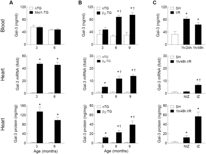
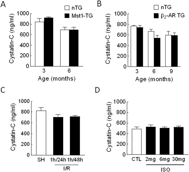
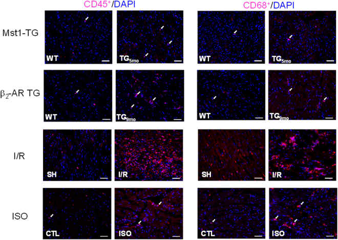
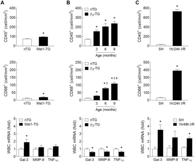
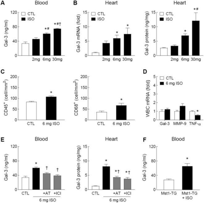
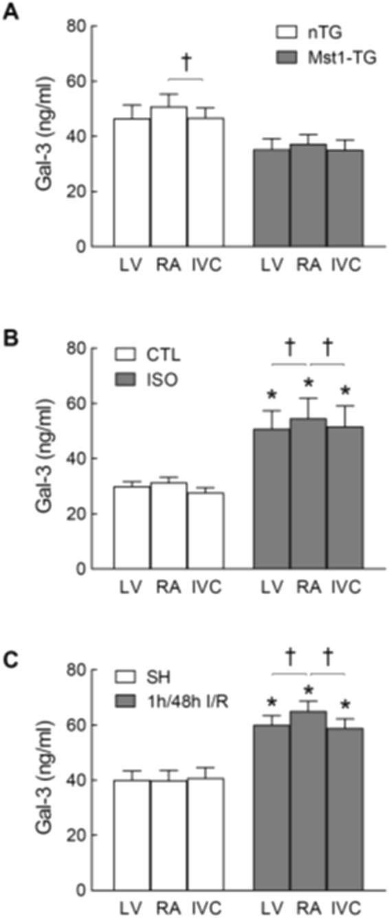
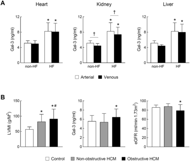
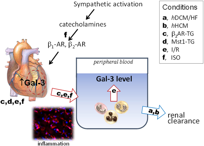
Galectin-3 is a biomarker of heart disease. However, it remains unknown whether increase in galectin-3 levels is dependent on aetiology or disease-associated conditions and whether diseased heart releases galectin-3 into the circulation. We explored these questions in mouse models of heart disease and in patients with cardiomyopathy. All mouse models (dilated cardiomyopathy, DCM; fibrotic cardiomyopathy, ischemia-reperfusion, I/R; treatment with β-adrenergic agonist isoproterenol) showed multi-fold increases in cardiac galectin-3 expression and preserved renal function. In mice with fibrotic cardiomyopathy, I/R or isoproterenol treatment, plasma galectin-3 levels and density of cardiac inflammatory cells were elevated. These models also exhibited parallel changes in cardiac and plasma galectin-3 levels and presence of trans-cardiac galectin-3 gradient, indicating cardiac release of galectin-3. DCM mice showed no change in circulating galectin-3 levels nor trans-cardiac galectin-3 gradient or myocardial inflammatory infiltration despite a 50-fold increase in cardiac galectin-3 content. In patients with hypertrophic cardiomyopathy or DCM, plasma galectin-3 increased only in those with renal dysfunction and a trans-cardiac galectin-3 gradient was not present. Collectively, this study documents the aetiology-dependency and diverse mechanisms of increment in circulating galectin-3 levels. Our findings highlight cardiac inflammation and enhanced β-adrenoceptor activation in mediating elevated galectin-3 levels via cardiac release in the mechanism.


Conflict of interest statement

The authors declare no competing interests.

Figures

Figure 1
Figure 1
Plasma and myocardial levels of galectin-3 in the mouse models of cardiomyopathy and ischemia-reperfusion (I/R). Changes in plasma concentrations of Gal-3 and cardiac expression of Gal-3 at mRNA and protein levels in Mst1-TG and non-TG (nTG) mice at 3 and 6 months of age (n = 6∼8/group, A) in β2-TG (n = 5∼9/group) and nTG mice (n = 4∼6/group) at 3, 6 and 9 months of age (B) and in mice subjected to 1-hour ischemia followed by reperfusion of 24 or 48 hours (n = 5∼7/group) or sham-operation as control (SH, n = 5, C). In hearts from mice subjected to I/R (1/48 h, n = 5) or SH (n = 4), the infarct zone (IZ) was separated from the non-infarct zone (NIZ) and the assays were performed separately. Mean ± SEM. *P < 0.05 vs. nTG or SH, and P < 0.05 vs. 3-month-old β2-TG or NIZ.
Figure 2
Figure 2
Plasma levels of the renal biomarker cyctatin-C in mouse models of heart disease. Mouse models consist of transgenic cardiomyopathy due to cardiac overexpression of Mst1 (A) or β2-AR (B) and mice subjected to ischemia/reperfusion (I/R, C) or treatment with isoproterenol (ISO, D). N = 5∼9 per group in all experiments.
Figure 3
Figure 3
Representative histological images of CD45 and CD68 cells by immunohistochemistry of the left ventricles. Images were from of two transgenic mouse models of cardiomyopathy, mice with ischemia/reperfusion (I/R, 1/48 h) or treated with isoproterenol (ISO, 6 mg/kg for 48 hours). Arrows indicate positively stained cells. Bar = 50 μm.
Figure 4
Figure 4
Inflammatory parameters of the myocardium and circulating leukocytes in mouse models of cardiomyopathy and ischemia-reperfusion (I/R). Cardiac and systemic inflammation was assessed by immunohistochemistry-based quantification of leukocytes (CD45+) or macrophages (CD68+) in the LV myocardium and by gene expression of Gal-3, matrix metalloproteinase-9 (MMP-9) and tumour necrosis factor-α (TNF-α) in circulating leukocytes. Results were from (A) Mst1-TG and nTG mice (n = 6/group for immunohistochemistry and n = 9/group for gene expression, 3–6 months of age). Data were combined from mice aged 3 and 6 months of age; (B) β2-TG and nTG mice aged at 3, 6 and 9 months (n = 5∼8 per age group for immunohistochemistry and n = 6∼12/group for gene expression of circulating leukocytes from 6-month-old mice); and (C) mice subjected to I/R (1/48 h) or sham-operation (SH) (n = 7/group for immunohistochemistry and n = 5∼8/group for gene expression). Mean ± SEM. *P < 0.05 vs. nTG or SH, P < 0.05 vs. 3-month-old β2-TG, #P < 0.05 vs. 6-month-old β2-TG.
Figure 5
Figure 5
Effect of the β-adrenoceptor agonist isoproterenol (ISO) on circulating and cardiac levels of galectin-3 and inflammatory parameters. Plasma levels of Gal-3 (A) and cardiac expression of Gal-3 (B) at the mRNA and protein levels in C57Bl/6 mice treated with the β-agonist ISO for 48 h at 2, 6 or 30 mg/kg/day (via subcutaneous osmotic minipump) or control without treatment (CTL). (C) Density of leukocytes (CD45+) or macrophages (CD68+) quantified by immunohistochemistry in hearts of mice without and with treatment of ISO at 6 mg/kg/day for 48 h. (D) Expression of Gal-3, matrix metalloproteinase-9 (MMP-9) and tumour necrosis factor-α (TNF-α) genes in circulating leukocytes from mice without and with ISO treatment (6 mg/kg/day for 48 hours). (E) ISO (6 mg/kg/day) stimulated Gal-3 upregulation was partially inhibited by atenolol (AT, β1-antagonist, 2 mg/kg/day) or by ICI-118551 (ICI, β2-antagonist, 1 mg/kg/day), both P < 0.05 vs. ISO alone. (F) Effect of treatment with ISO (6 mg/kg/day for 48 hours) on circulating level of Gal-3 in Mst1-TG mice. *P < 0.05 vs. control (CTL) or untreated Mst1-TG, #P < 0.05 vs. 2 mg/kg/day ISO group, and P < 0.05 vs. 6 mg/kg/day ISO group. Mean ± SEM. N = 5∼8/group in all experiments.
Figure 6
Figure 6
Galectin-3 gradients of plasma samples from three sites of the heart in different mouse models. In each mouse, blood was sampled from the right atrium (RA), left ventricle (LV) and inferior vena cava (IVC), respectively. Blood was collected from Mst1-TG and nTG mice (n = 9 and 6, respectively; A) C57Bl/6 mice treated with isoproterenol (ISO, 6 mg/kg/day for 48 h, n = 9) untreated control (CTL, n = 6, B) or subjected to ischemia/reperfusion (I/R, 1/48 h, n = 8) or sham-operation (SH, n = 5, C). Mean ± SEM. *P < 0.05 vs. CTL, SH or nTG, respectively, by two-way ANOVA. P < 0.05 by paired t-test.
Figure 7
Figure 7
Plasma levels of galectin-3 in cardiomyopathy patients with and without heart failure. (A) Arterial and venous plasma levels of Gal-3 across the heart, kidney and liver from cardiomyopathy patients with HF (n = 15) and non-HF subjects (n = 5). Arterial blood was collected from the radial or brachial artery. Venous blood across the heart, kidney and liver was collected from the coronary sinus, renal vein and hepatic vein, respectively. Paired t-test was used to determine trans-organ effect of plasma Gal-3. Mean ± SD. *P < 0.05 vs. non-HF control. P < 0.05 by paired-t-test. (B) Left ventricular mass index (LVMI), plasma level of Gal-3 and estimated glomerular filtration rate (eGFR) in healthy controls (n = 20) and patients with non-obstructive (n = 27) or obstructive (n = 21) hypertrophic cardiomyopathy (HCM). Mean ± SD. *P < 0.05 vs. control group and #P < 0.05 vs. non-obstructive HCM group.
Figure 8
Figure 8
Determinants of elevated circulating levels of galectin-3 in cardiomyopathy and heart disease. Changes in circulating Gal-3 level is dependent on the aetiology of cardiomyopathy, inflammatory status and renal function. While cardiac release of Gal-3 is not observed in Mst1-TG DCM mice and HF patients (hDCM for human DCM, hHCM for human HCM), this mechanism is strongly indicated by findings from animal models of β2-TG cardiomyopathy, ischemia/reperfusion (I/R) or treatment with isoproterenol (ISO). Two factors closely associated with heart disease, i.e. cardiac and/or systemic inflammation and activation of the sympatho-β-adrenergic system, contribute cardiac Gal-3 release and increased blood levels of Gal-3. Red arrows indicate potential sources whilst blue arrow denotes renal clearance of circulating Gal-3 pool.

References

    1. Rabinovich GA, Toscano MA. Turning ‘sweet’ on immunity: galectin-glycan interactions in immune tolerance and inflammation. Nat Rev Immunol. 2009;9:338–52. doi: 10.1038/nri2536. - DOI - PubMed
    1. van der Velde AR, et al. Prognostic value of changes in galectin-3 levels over time in patients with heart failure: data from CORONA and COACH. Circ Heart Fail. 2013;6:219–226. doi: 10.1161/CIRCHEARTFAILURE.112.000129. - DOI - PubMed
    1. Clementy N, et al. Serum Galectin-3 Levels Predict Recurrences after Ablation of Atrial Fibrillation. Sci Rep. 2016;6:34357. doi: 10.1038/srep34357. - DOI - PMC - PubMed
    1. Francia P, et al. Osteopontin and galectin-3 predict the risk of ventricular tachycardia and fibrillation in heart failure patients with implantable defibrillators. J Cardiovasc Electrophysiol. 2014;25:609–616. doi: 10.1111/jce.12364. - DOI - PubMed
    1. Hernandez-Romero D, et al. Galectin-3 as a marker of interstitial atrial remodelling involved in atrial fibrillation. Sci Rep. 2017;7:40378. doi: 10.1038/srep40378. - DOI - PMC - PubMed

Publication types