. 2018 May 29;9(1):2105.
doi: 10.1038/s41467-018-04521-0.

Dominant-negative STAT5B mutations cause growth hormone insensitivity with short stature and mild immune dysregulation

Affiliations

Dominant-negative STAT5B mutations cause growth hormone insensitivity with short stature and mild immune dysregulation

Jürgen Klammt et al. Nat Commun. .

Abstract

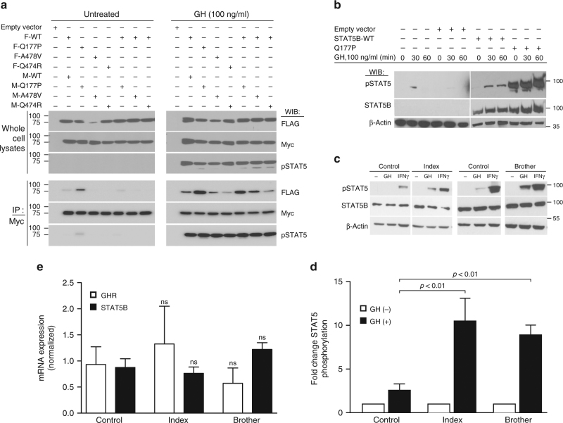
Growth hormone (GH) insensitivity syndrome (GHIS) is a rare clinical condition in which production of insulin-like growth factor 1 is blunted and, consequently, postnatal growth impaired. Autosomal-recessive mutations in signal transducer and activator of transcription (STAT5B), the key signal transducer for GH, cause severe GHIS with additional characteristics of immune and, often fatal, pulmonary complications. Here we report dominant-negative, inactivating STAT5B germline mutations in patients with growth failure, eczema, and elevated IgE but without severe immune and pulmonary problems. These STAT5B missense mutants are robustly tyrosine phosphorylated upon stimulation, but are unable to nuclear localize, or fail to bind canonical STAT5B DNA response elements. Importantly, each variant retains the ability to dimerize with wild-type STAT5B, disrupting the normal transcriptional functions of wild-type STAT5B. We conclude that these STAT5B variants exert dominant-negative effects through distinct pathomechanisms, manifesting in milder clinical GHIS with general sparing of the immune system.


Conflict of interest statement

R.G.R. consults for OPKO, Versartis, Ascendis, Genexine, Ammonite, Sandoz, Ferring, and NovoNordisk. The remaining authors declare no competing interests.

Figures

Fig. 1
Fig. 1
Segregation of heterozygous STAT5B missense mutations in the families. a Individuals bearing de novo (family 1) or inherited (families 2 and 3) heterozygous STAT5B mutations are indicated by half-filled symbols. Standard deviation score of last reported height [SDS], relative immunoglobulin E [IgE] levels, and the occurrence of eczema are shown below the symbols. Index patients are marked by an arrow. Question marks indicate an unknown genotype. n.d., no data available. b Schematic of the STAT5B protein (drawn to scale) with major functional domains (domains: ND, N-terminal; CCD, coiled-coil; DBD, DNA-binding; L, linker; SH2, src-homology 2; TAD, transactivation), their boundaries, and tyrosine residue (Tyr699) whose phosphorylation is necessary for STAT5B activation. Positions of previously reported homozygous amino acid substitutions are shown above the bar (arrows: red, protein truncating variants; blue, missense); heterozygous missense mutations identified in this study are indicated below the bar (green arrows)
Fig. 2
Fig. 2
GH-dependent STAT5B activation is retained by the missense variants. Whole-cell lysates from untreated or GH-stimulated (30 min or as shown) HEK293(hGHR) (a) or HEK293 (b) cells transfected with the indicated FLAG- (F) or Myc-tagged (M) STAT5B wild-type (WT) or variant plasmids (p.Gln177Pro, Q177P; p.Ala478Val, A478V; p.Gln474R, Q474R) were subjected to immunoprecipitation (IP) and/or immunoblotting (WIB) using antibodies as indicated (pSTAT5, phospho-Tyr-STAT5). Representative immunoblots of three experiments are shown. c–e Whole-cell lysates were prepared from primary dermal fibroblasts derived from affected twins in Family 1 and control fibroblasts, after stimulation with GH or IFNγ and immunoblotted for pSTAT5, total STAT5B, or β-actin as a loading control. Representative blot out of four independent experiments is shown. d Densitometric evaluation of pSTAT5 determined in patients’ fibroblasts (fold change compared to untreated cells). pSTAT5 levels were normalized to total STAT5B and β-actin protein amounts in each sample. Data are shown as mean ± S.E.M. of three independent experiments. e qPCR verification of STAT5B and GHR mRNA expression determined in fibroblasts from the index patient, his brother, and control cells. Mean ± S.E.M. of at least three independent experiments is shown. Statistical analysis was performed by one-way ANOVA followed by Tukey’s post hoc test, p-values relative to control are indicated (ns, not significant)
Fig. 3
Fig. 3
STAT5B missense variants are defective in nuclear translocation or DNA binding. a Representative immunofluorescence images of untreated or GH-stimulated HEK293(hGHR) cells transfected with Myc-STAT5B wild type (WT) or Myc-STAT5B variant plasmids (as indicated, abbreviations as in Fig. 2) and stained with α-Myc antibodies and Hoechst 33342 nuclear staining (scale bar, 10 µm). b Images of GH-stimulated or untreated HEK293(hGHR) cells transiently transfected with Myc-STAT5B wild type (WT) and co-transfected with plasmids expressing WT (upper panel) or p.Gln177Pro STAT5B (Q177P, bottom panel) followed by immunostaining (IF) with α-FLAG or α-Myc antibodies and Hoechst 33342 (scale bar, 10 µm). c, d Representative (three independent experiments) electrophoretic mobility shift assays (EMSA) (c) or supershift EMSA (d) to demonstrate DNA-binding of STAT5B WT and/or variant proteins as indicated. d Supershift EMSA of FLAG-WT co-transfected with indicated Myc-tagged WT or variants (V, vector only control; arrow, shifted; arrowhead, supershifted complexes). e Luciferase reporter activity measured in HEK293(hGHR) cells co-transfected with equimolar amounts of the indicated FLAG-tagged plasmids. Activities are presented as fold increase above unstimulated vector control, which was arbitrarily assigned a value of 1.0. Error bars represent mean ± S.D. of four independent experiments, each time in duplicates. *p < 0.05, **p < 0.01 relative to GH-stimulated WT, using one-way ANOVA with post hoc Tukey test

References

    1. David A, et al. Evidence for a continuum of genetic, phenotypic, and biochemical abnormalities in children with growth hormone insensitivity. Endocr. Rev. 2011;32:472–497. doi: 10.1210/er.2010-0023. - DOI - PubMed
    1. Rosenfeld RG, Rosenbloom AL, Guevara-Aguirre J. Growth hormone (GH) insensitivity due to primary GH receptor deficiency. Endocr. Rev. 1994;15:369–390. doi: 10.1210/edrv-15-3-369. - DOI - PubMed
    1. Laron Z. Laron syndrome (primary growth hormone resistance or insensitivity): the personal experience 1958-2003. J. Clin. Endocrinol. Metab. 2004;89:1031–1044. doi: 10.1210/jc.2003-031033. - DOI - PubMed
    1. Ayling RM, et al. A dominant-negative mutation of the growth hormone receptor causes familial short stature. Nat. Genet. 1997;16:13–14. doi: 10.1038/ng0597-13. - DOI - PubMed
    1. Iida K, et al. Growth hormone (GH) insensitivity syndrome with high serum GH-binding protein levels caused by a heterozygous splice site mutation of the GH receptor gene producing a lack of intracellular domain. J. Clin. Endocrinol. Metab. 1998;83:531–537. - PubMed

Publication types