. 2018 May 29;8(1):8222.
doi: 10.1038/s41598-018-26584-1.

Expansion of human primary hepatocytes in vitro through their amplification as liver progenitors in a 3D organoid system

Affiliations

Expansion of human primary hepatocytes in vitro through their amplification as liver progenitors in a 3D organoid system

Delphine Garnier et al. Sci Rep. .

Abstract

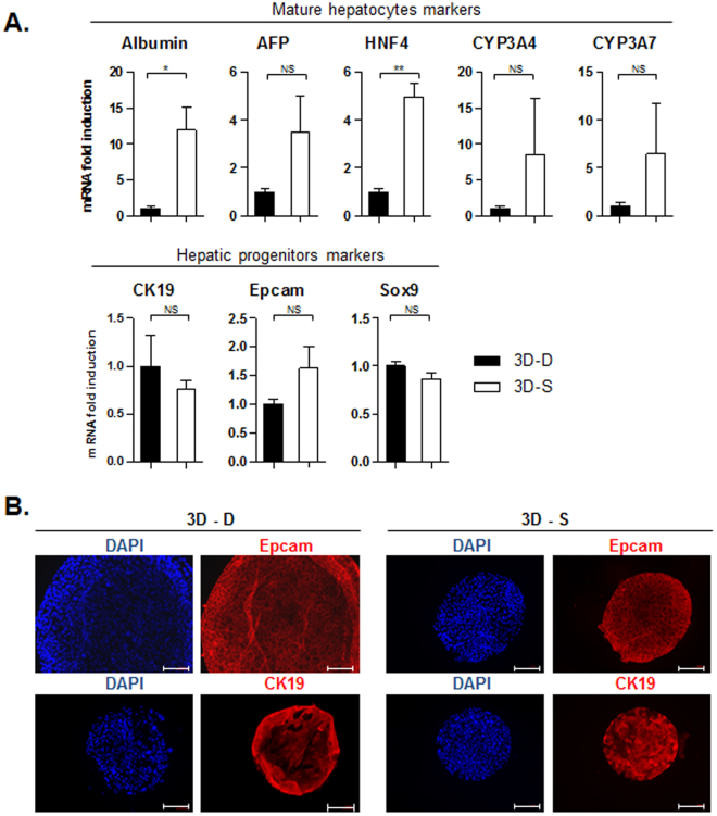
Despite decades of investigation on the proliferation of adult human primary hepatocytes, their expansion in vitro still remains challenging. To later be able to consider hepatocytes as a cell therapy alternative or bridge to liver transplantation, dramatically impeded by a shortage in liver donors, the first step is having an almost unlimited source of these cells. The banking of transplantable hepatocytes also implies a protocol for their expansion that can be compatible with large-scale production. We show that adult human primary hepatocytes when grown in 3D organoids are easily amplified, providing a substantial source of functional hepatocytes ready for transplantation. Following their plating, differentiated human hepatocytes are amplified during a transient and reversible step as liver progenitors, and can subsequently be converted back to mature differentiated hepatocytes. The protocol we propose is not only compatible with automated and high-throughput cell culture systems, thanks to the expansion of hepatocytes in suspension, but also guarantees the generation of a high number of functional cells from the same patient sample, with a relatively easy set up.


Conflict of interest statement

The authors declare no competing interests.

Figures

Figure 1
Figure 1
Variability in kinetic of growth of human hepatocytes when cultured as 3D organoids, depending on the cell batch. (A) Characteristics of human hepatocytes batches used. (B) Top panel shows organoids counting per well, 1 and 2 weeks after plating. Middle panel indicates the cell number production per well of a 24-wells plate over time. Cells were dissociated and counted once a week, and plated back in the same conditions. And the respective percentage of Epcam positive cells in each batch, measured by FACS, is shown in lower panel.
Figure 2
Figure 2
Culture of human hepatocytes as 3D organoids leads to long term survival, loss of mature hepatocyte markers, and enrichment in liver progenitors markers. (A) The expression of mature hepatocytes markers was analyzed by qPCR in adherent hepatocytes at 24, 48 and 72 h after plating (Adh), or in hepatocyte organoids (3D) after 2–3 passages (t1; around 40 days after plating) and 2 weeks later (t2). (B) The expression of markers of liver progenitors was analyzed in the same conditions, as well as Ki67 proliferation marker (C). The expression was normalized compared to the expression in adherent human hepatocytes 24 h post-plating. (N = 3).
Figure 3
Figure 3
3D organoid culture of mature human hepatocytes in Matrigel drops or in Matrigel suspension display comparable levels of viability and growth on short-term. (A) Observation by phase contrast microscopy of human hepatocytes cultured as adherent cells, in suspension, or maintained in 3D inside Matrigel drop or in Matrigel suspension (bar graph = 50 μm). (B) Schematic representation of the protocol settings. (C) Average organoid counts 3 weeks after plating as 3D, in Matrigel drops (3D-D - black bars) or in Matrigel suspension (3D-S - white bars) (number of organoids per well of a 24-wells plate) (N = 2). (D) Analysis of cell viability with Live (green)/Dead (red) staining kit (bar graph = 50 μm). Proliferation was analyzed by Ki67 immunofluorescence (E) (bar graph = 100 μm) and qPCR (F) (N = 3).
Figure 4
Figure 4
3D organoids cultured in Matrigel suspension also allows amplification of liver progenitors, while maintaining a higher level of differentiated functions. (A) The expression of different liver progenitors/mature hepatocyte/proliferation markers was analyzed at the mRNA level by qPCR, in 3D culture of human hepatocytes inside Matrigel drop (3D-D - black bars) or in suspension (3D-S - white bars), after 2–3 passages (t1; close to 40 days after plating) and 2 weeks later (t2). Data were normalized to the expression in Matrigel drop organoids. (N = 3) (B) The expression of some of those markers was validated at the protein level by immunofluorescence (bar graph = 100 μm).
Figure 5
Figure 5
Induction of differentiation of 3D organoids into mature hepatocytes is more efficient if cultured in Matrigel suspension. The mRNA expression level of different liver progenitor/mature hepatocyte/proliferation markers was analyzed by qPCR, in 3D culture of human hepatocytes inside Matrigel drops (3D-D – black bars) or in Matrigel suspension (3D-S – white bars), in expansion (EM) or differentiation media (DM). (N = 3).

References

    1. Michalopoulos GK. Hepatostat: Liver regeneration and normal liver tissue maintenance. Hepatology. 2017;65:1384–1392. doi: 10.1002/hep.28988. - DOI - PubMed
    1. Michalopoulos GK, Khan Z. Liver Stem Cells: Experimental Findings and Implications for Human Liver Disease. Gastroenterology. 2015;149:876–882. doi: 10.1053/j.gastro.2015.08.004. - DOI - PMC - PubMed
    1. Fausto N, Campbell JS. The role of hepatocytes and oval cells in liver regeneration and repopulation. Mechanisms of Development. 2003;120:117–130. doi: 10.1016/S0925-4773(02)00338-6. - DOI - PubMed
    1. Kopp JL, Grompe M, Sander M. Stem cells versus plasticity in liver and pancreas regeneration. Nat. Cell Biol. 2016;18:238–245. doi: 10.1038/ncb3309. - DOI - PubMed
    1. Miyajima A, Tanaka M, Itoh T. Stem/progenitor cells in liver development, homeostasis, regeneration, and reprogramming. Cell Stem Cell. 2014;14:561–574. doi: 10.1016/j.stem.2014.04.010. - DOI - PubMed

Publication types