Review
. 2018 Sep;15(9):555-567.
doi: 10.1038/s41575-018-0020-y.

Liver sinusoidal endothelial cells - gatekeepers of hepatic immunity

Affiliations
Review

Liver sinusoidal endothelial cells - gatekeepers of hepatic immunity

Shishir Shetty et al. Nat Rev Gastroenterol Hepatol. 2018 Sep.

Abstract

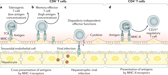
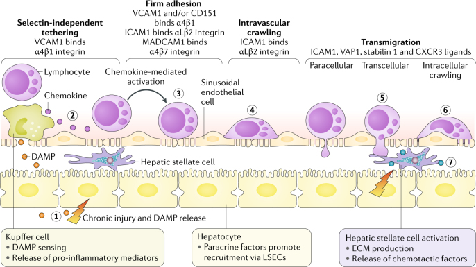
Liver sinusoidal endothelial cells (LSECs) line the low shear, sinusoidal capillary channels of the liver and are the most abundant non-parenchymal hepatic cell population. LSECs do not simply form a barrier within the hepatic sinusoids but have vital physiological and immunological functions, including filtration, endocytosis, antigen presentation and leukocyte recruitment. Reflecting these multifunctional properties, LSECs display unique structural and phenotypic features that differentiate them from the capillary endothelium present within other organs. It is now clear that LSECs have a critical role in maintaining immune homeostasis within the liver and in mediating the immune response during acute and chronic liver injury. In this Review, we outline how LSECs influence the immune microenvironment within the liver and discuss their contribution to immune-mediated liver diseases and the complications of fibrosis and carcinogenesis.


Conflict of interest statement

The authors have no competing financial interests.

Figures

Fig. 1
Fig. 1. Microanatomy of the human liver vascular tree.
a | Low-power image of human liver tissue (stained with haematoxylin and eosin) illustrating the lobular organization of the liver, with zonal architecture indicated relative to the position of the portal tract. b | Expanded periportal section of the same image to illustrate the different vascular compartments within the parenchyma. c | Immunohistochemical staining of stabilin 1, which highlights liver endothelial cell distribution within hepatic tissue in a normal liver section. d | A comparison of the structure of liver sinusoidal endothelium and glomerular endothelium.
Fig. 2
Fig. 2. Hepatic sinusoidal endothelial cells as antigen-presenting cells.
a | Liver sinusoidal endothelial cells (LSECs) express major histocompatibility complex class I (MHC-I) receptors and can cross-present antigen to CD8+ cytotoxic T cells. At low antigen concentrations, this presentation leads to tolerance and deletion of CD8+ T cells. b | If antigen concentrations are high, then antigen cross presentation to CD8+ T cells leads to a memory effector T cell phenotype. c | In the context of hepatotrophic infections such as hepatitis B, CD8+ T cells adhere to the sinusoids in a platelet-dependent process and then probe for infected hepatocytes through LSEC fenestrae. Detection of an infected hepatocyte leads to diapedesis (the process of cells actively crossing capillaries)-independent killing. d | LSECs can also present antigen to CD4+ T cells via expression of MHC class II, which leads to the induction of suppressor T cells (CD25hi regulatory T cells). TCR, T cell receptor.
Fig. 3
Fig. 3. Lymphocyte recruitment within the hepatic sinusoids.
Lymphocyte recruitment involves an adhesion cascade within the hepatic sinusoids that is influenced by the low shear environment and cellular crosstalk between parenchymal and non-parenchymal cells. Chronic parenchymal cell damage leads to the release of danger-associated molecular patterns (DAMPs) and pro-inflammatory mediators by Kupffer cells, which increase adhesion molecule expression by liver sinusoidal endothelial cells (LSECs) (step 1). Lymphocyte recruitment across activated LSECs involves a selectin-independent tethering step (step 2), followed by integrin activation and firm adhesion to immunoglobulin superfamily members on the LSEC surface (step 3). This process is influenced by paracrine factors released from hepatocytes. Lymphocytes then crawl along the luminal endothelium (step 4) until they receive a signal to transmigrate across LSECs through either a paracellular or a transcellular route (step 5). A third route of lymphocyte migration involves intracellular migration directly into the LSEC body and then migration to the adjacent LSEC, termed intracellular crawling (step 6). Release of chemotactic factors from activated hepatic stellate cells promotes subsequent migration and positioning in liver tissue (step 7). CXCR3, CXC-chemokine receptor 3; ECM, extracellular matrix; ICAM1, intercellular adhesion molecule 1; MADCAM1, mucosal addressin cell adhesion molecule 1; VAP1, vascular adhesion protein1; VCAM1, vascular cell adhesion molecule 1.

References

    1. Sorensen KK, Simon-Santamaria J, McCuskey RS, Smedsrod B. Liver sinusoidal endothelial cells. Compr. Physiol. 2015;5:1751–1774. doi: 10.1002/cphy.c140078. - DOI - PubMed
    1. Poisson J, et al. Liver sinusoidal endothelial cells: physiology and role in liver diseases. J. Hepatol. 2017;66:212–227. doi: 10.1016/j.jhep.2016.07.009. - DOI - PubMed
    1. Mates JM, et al. Mouse liver sinusoidal endothelium eliminates HIV-like particles from blood at a rate of 100 million per minute by a second-order kinetic process. Front. Immunol. 2017;8:35. doi: 10.3389/fimmu.2017.00035. - DOI - PMC - PubMed
    1. Smedsrod B. Clearance function of scavenger endothelial cells. Comp. Hepatol. 2004;3(Suppl. 1):S22. doi: 10.1186/1476-5926-2-S1-S22. - DOI - PMC - PubMed
    1. Li R, et al. Role of liver sinusoidal endothelial cells and stabilins in elimination of oxidized low-density lipoproteins. Am. J. Physiol. Gastrointest. Liver Physiol. 2011;300:G71–G81. doi: 10.1152/ajpgi.00215.2010. - DOI - PMC - PubMed

Publication types