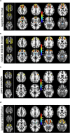
The anatomy of the human medial forebrain bundle: Ventral tegmental area connections to reward-associated subcortical and frontal lobe regions
- PMID: 29845013
- PMCID: PMC5964495
- DOI: 10.1016/j.nicl.2018.03.019
The anatomy of the human medial forebrain bundle: Ventral tegmental area connections to reward-associated subcortical and frontal lobe regions
Abstract
Introduction: Despite their importance in reward, motivation, and learning there is only sparse anatomical knowledge about the human medial forebrain bundle (MFB) and the connectivity of the ventral tegmental area (VTA). A thorough anatomical and microstructural description of the reward related PFC/OFC regions and their connection to the VTA - the superolateral branch of the MFB (slMFB) - is however mandatory to enable an interpretation of distinct therapeutic effects from different interventional treatment modalities in neuropsychiatric disorders (DBS, TMS etc.). This work aims at a normative description of the human MFB (and more detailed the slMFB) anatomy with respect to distant prefrontal connections and microstructural features.
Methods and material: Healthy subjects (n = 55; mean age ± SD, 40 ± 10 years; 32 females) underwent high resolution anatomical magnetic resonance imaging including diffusion tensor imaging. Connectivity of the VTA and the resulting slMFB were investigated on the group level using a global tractography approach. The Desikan/Killiany parceling (8 segments) of the prefrontal cortex was used to describe sub-segments of the MFB. A qualitative overlap with Brodmann areas was additionally described. Additionally, a pure visual analysis was performed comparing local and global tracking approaches for their ability to fully visualize the slMFB.
Results: The MFB could be robustly described both in the present sample as well as in additional control analyses in data from the human connectome project. Most VTA- connections reached the superior frontal gyrus, the middel frontal gyrus and the lateral orbitofrontal region corresponding to Brodmann areas 10, 9, 8, 11, and 11m. The projections to these regions comprised 97% (right) and 98% (left) of the total relative fiber counts of the slMFB.
Discussion: The anatomical description of the human MFB shows far reaching connectivity of VTA to reward-related subcortical and cortical prefrontal regions - but not to emotion-related regions on the medial cortical surface - realized via the superolateral branch of the MFB. Local tractography approaches appear to be inferior in showing these far-reaching projections. Since these local approaches are typically used for surgical targeting of DBS procedures, the here established detailed map might - as a normative template - guide future efforts to target deep brain stimulation of the slMFB in depression and other disorders related to dysfunction of reward and reward-associated learning.
Keywords: Brain; Deep brain stimulation; Depression; Human; Medial forebrain bundle; Normal anatomy; Obsessive compulsive disorder; TMS.
Figures


Similar articles
-
Joint Anatomical, Histological, and Imaging Investigation of the Midbrain Target Region for Superolateral Medial Forebrain Bundle Deep Brain Stimulation.Stereotact Funct Neurosurg. 2025;103(1):1-13. doi: 10.1159/000541834. Epub 2024 Nov 11. Stereotact Funct Neurosurg. 2025. PMID: 39527932 Free PMC article.
-
Tractography-assisted deep brain stimulation of the superolateral branch of the medial forebrain bundle (slMFB DBS) in major depression.Neuroimage Clin. 2018 Aug 14;20:580-593. doi: 10.1016/j.nicl.2018.08.020. eCollection 2018. Neuroimage Clin. 2018. PMID: 30186762 Free PMC article. Clinical Trial.
-
Ventral tegmental area connections to motor and sensory cortical fields in humans.Brain Struct Funct. 2019 Nov;224(8):2839-2855. doi: 10.1007/s00429-019-01939-0. Epub 2019 Aug 22. Brain Struct Funct. 2019. PMID: 31440906 Free PMC article.
-
The medial forebrain bundle as a deep brain stimulation target for treatment resistant depression: A review of published data.Prog Neuropsychopharmacol Biol Psychiatry. 2015 Apr 3;58:59-70. doi: 10.1016/j.pnpbp.2014.12.003. Epub 2014 Dec 19. Prog Neuropsychopharmacol Biol Psychiatry. 2015. PMID: 25530019 Review.
-
Electrical stimulation of the medial forebrain bundle in pre-clinical studies of psychiatric disorders.Neurosci Biobehav Rev. 2015 Feb;49:32-42. doi: 10.1016/j.neubiorev.2014.11.018. Epub 2014 Dec 9. Neurosci Biobehav Rev. 2015. PMID: 25498857 Review.
Cited by
-
The Neuroanatomy of the Habenular Complex and Its Role in the Regulation of Affective Behaviors.J Funct Morphol Kinesiol. 2024 Jan 3;9(1):14. doi: 10.3390/jfmk9010014. J Funct Morphol Kinesiol. 2024. PMID: 38249091 Free PMC article. Review.
-
Deep brain stimulation of the medial forebrain bundle for treatment-resistant depression - a narrative literature review.Postep Psychiatr Neurol. 2021 Sep;30(3):183-189. doi: 10.5114/ppn.2021.110788. Epub 2021 Nov 26. Postep Psychiatr Neurol. 2021. PMID: 37082768 Free PMC article. Review.
-
Microstructural Changes in the Left Mesocorticolimbic Pathway are Associated with the Comorbid Development of Fatigue and Depression in Multiple Sclerosis.J Neuroimaging. 2021 May;31(3):501-507. doi: 10.1111/jon.12832. Epub 2021 Feb 1. J Neuroimaging. 2021. PMID: 33522683 Free PMC article.
-
The rostro-caudal gradient in the prefrontal cortex and its modulation by subthalamic deep brain stimulation in Parkinson's disease.Sci Rep. 2021 Jan 22;11(1):2138. doi: 10.1038/s41598-021-81535-7. Sci Rep. 2021. PMID: 33483554 Free PMC article.
-
Deep brain stimulation of the "medial forebrain bundle": a strategy to modulate the reward system and manage treatment-resistant depression.Mol Psychiatry. 2022 Jan;27(1):574-592. doi: 10.1038/s41380-021-01100-6. Epub 2021 Apr 26. Mol Psychiatry. 2022. PMID: 33903731 Review.
References
-
- Andersson J.L.R., Skare S., Ashburner J. How to correct susceptibility distortions in spin-echo echo-planar images: application to diffusion tensor imaging. NeuroImage. 2003 Oct;20(2):870–888. - PubMed
-
- Anthofer J.M., Steib K., Fellner C., Lange M., Brawanski A., Schlaier J. DTI-based deterministic fibre tracking of the medial forebrain bundle. Acta Neurochir. Springer Vienna. 2015 Mar;157(3):469–477. - PubMed
-
- BECK E. The origin, course and termination of the prefronto-pontine tract in the human brain. Brain. 1950 Jan 1;73(3):368–391. - PubMed
-
- Beck A.T., Steer R.A., Ball R., Ranieri W. Comparison of beck depression inventories -IA and -II in psychiatric outpatients. J. Pers. Assess. 1996 Dec;67(3):588–597. - PubMed
Publication types
MeSH terms
LinkOut - more resources
Full Text Sources
Other Literature Sources
Miscellaneous

