. 2018 Oct;41(10):1530-1544.
doi: 10.1007/s00270-018-2000-6. Epub 2018 May 29.

Microwave Ablation in the Management of Colorectal Cancer Pulmonary Metastases

Affiliations

Microwave Ablation in the Management of Colorectal Cancer Pulmonary Metastases

I Kurilova et al. Cardiovasc Intervent Radiol. 2018 Oct.

Abstract

Purpose: To review outcomes following microwave ablation (MWA) of colorectal cancer pulmonary metastases and assess predictors of oncologic outcomes.

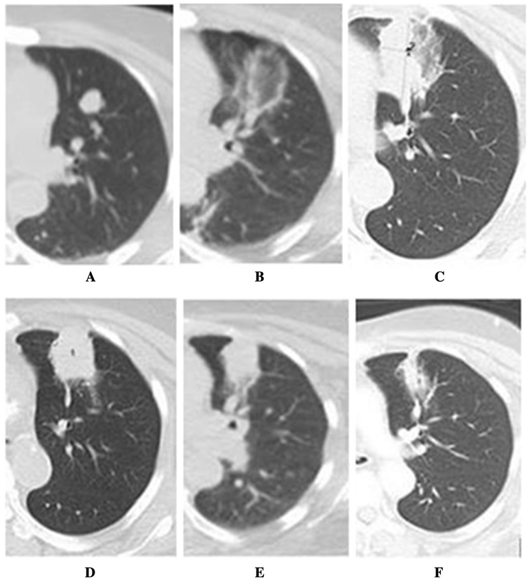
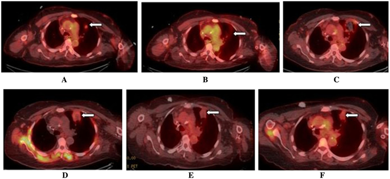
Methods: Technical success, primary and secondary technique efficacy rates were evaluated for 50 patients with 90 colorectal cancer pulmonary metastases at immediate, 4-8 weeks post-MWA and subsequent follow-up CT and/or 18F-FDG PET/CT. Local tumor progression (LTP) rate, LTP-free survival (LTPFS), cancer-specific and overall survivals were assessed. Complications were recorded according to SIR classification.

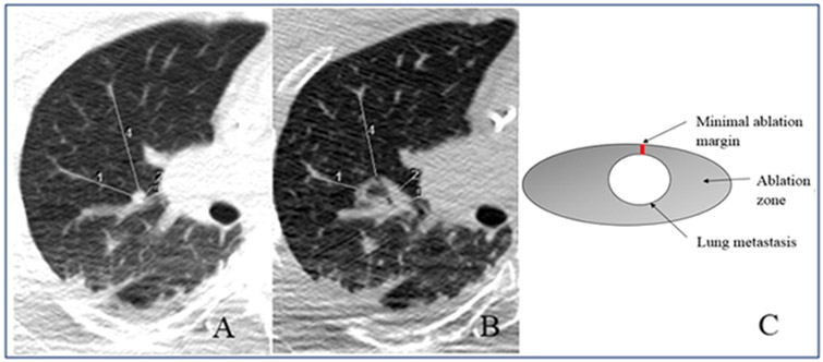
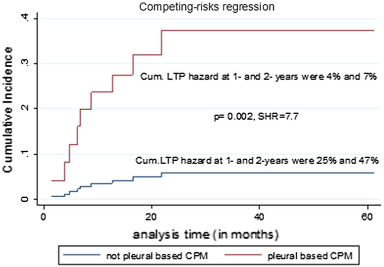
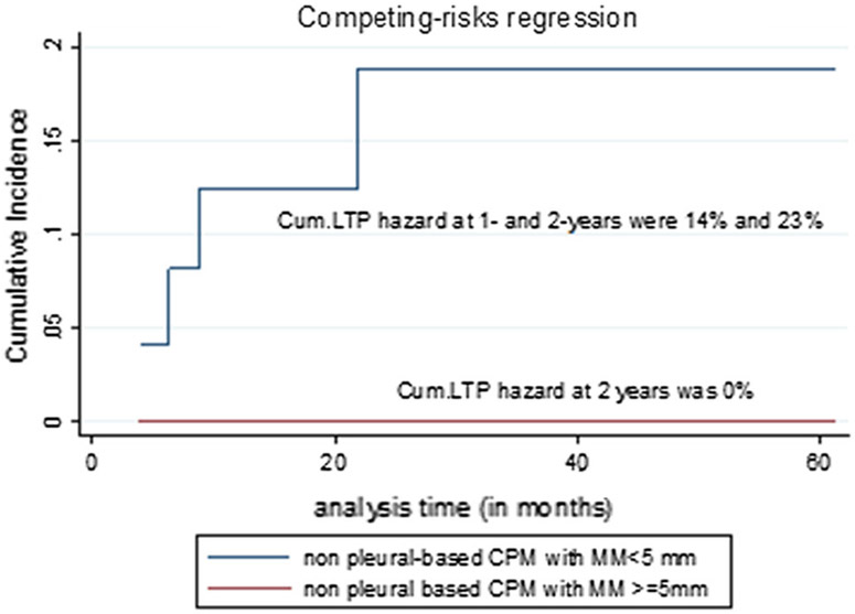
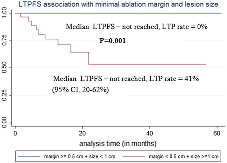
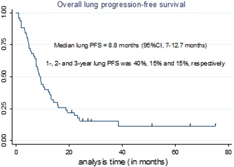
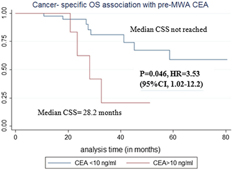
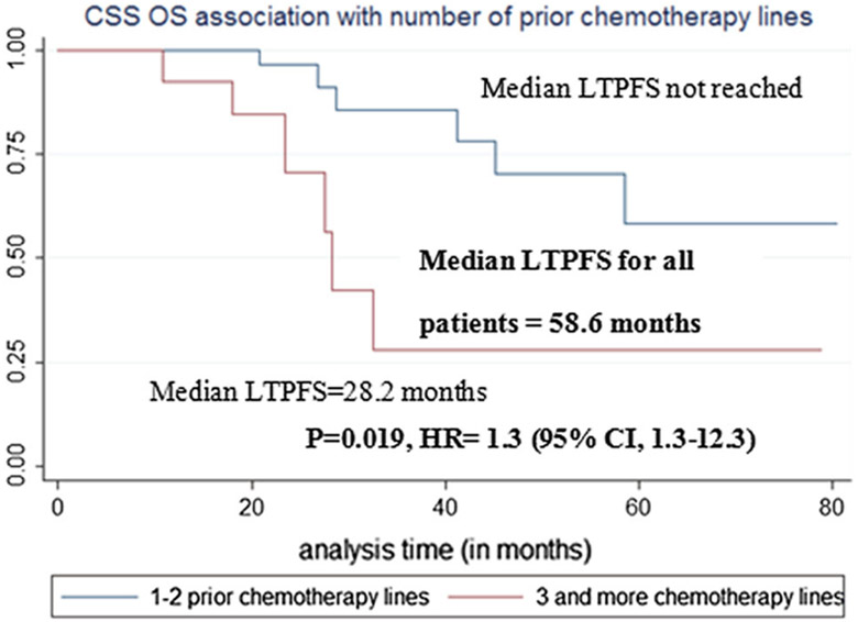
Results: Median follow-up was 25.6 months. Median tumor size was 1 cm (0.3-3.2 cm). Technical success, primary and secondary technique efficacy rates were 99, 90 and 92%, respectively. LTP rate was 10%. One-, 2- and 3-year LTPFS were: 93, 86 and 86%, respectively, with median LTPFS not reached. Median overall survival was 58.6 months, and median cancer-specific survival (CSS) was not reached. One-, 2- and 3-year overall and CSS were 94% and 98, 82 and 90%, 61 and 70%, respectively. On univariate analysis, minimal ablation margin (p < 0.001) and tumor size (p = 0.001) predicted LTPFS, with no LTP for minimal margin ≥ 5 mm and/or tumor size < 1 cm. Pleural-based metastases were associated with increased LTP risk (p = 0.002, SHR = 7.7). Pre-MWA CEA level > 10 ng/ml (p = 0.046) and ≥ 3 prior chemotherapy lines predicted decreased CSS (p = 0.02). There was no 90-day death. Major complications rate was 13%.

Conclusions: MWA with minimal ablation margin ≥ 5 mm is essential for local control of colorectal cancer pulmonary metastases. Pleural-based metastases and larger tumor size were associated with higher risk of LTP. CEA level and pre-MWA chemotherapy impacted CSS.

Keywords: Colorectal cancer; Lung ablation; Microwave ablation; Pulmonary metastases; Thermal ablation.


Conflict of interest statement

Conflict of interest C.T. Sofocleous has received research support from BTG, Ethicon (Neuwave); HS Medical, Angiodynamics; Sota Medical; and is a consultant for Ethicon and GE. S.B. Solomon, shareholder of Johnson & Johnson, has received personal fees from Medtronics, Astra Zeneca, Johnson & Johnson and GE Heathcare. Other authors have no conflict of interest.

Figures

Fig. 1
Fig. 1
Minimal ablation margin measurement. A pre-ablation measurements: measurement (1) = 34 mm; measurement (2) = 14 mm; measurement (3) = 8 mm; measurement (4) = 50 mm. B Post-ablation measurements: measurement (1) = 19 mm; measurement (2) = 12 mm; measurement (3) = 3 mm; measurement (4) = 37 mm. After subtracting corresponding measurements at each location [1-4], the respective ablation margins were 15, 2, 5 and 13 mm. Therefore, minimal ablation margin for this ablation zone was 2 mm (insufficient). C Minimal ablation margin scheme
Fig. 2
Fig. 2
Lung MWA ablation zone changes in 72-year old woman with metastatic colorectal cancer over 28-month follow-up period on cross-sectional CT, showing gradual constriction of ablation zone and no evidence of local tumor progression. After initial diagnosis, the patient underwent right hemicolectomy, adjuvant chemotherapy with 5-FU and leucovorin and was off-treatment with no evidence of disease for 18 months. A Pre-ablation enlarging lung nodule (from 0.9 to 1.6 cm) with rising CEA (from 5.8 to 11.6 ng/ml): lesion was considered metastatic, not biopsy-proven. Due to significant comorbidities (end-stage kidney insufficiency, coronary artery disease, diabetes, arterial hypertension and history of stroke), lung ablation was preferred to surgery. B Immediate post-MWA CT with ablation zone as ground glass opacity measuring 3.2 × 3.0 cm. C First post-MWA scan at 5 weeks, served as a new baseline for future comparisons. D Follow-up scan at 17 weeks. E Follow-up scan at 15.5 months; F follow-up scan at 28 months with constricted ablation zone and no evidence of local tumor progression. The patient was off-treatment after MWA with no evidence of disease elsewhere for 2 years, when the patient developed solitary biopsy-proven liver metastasis, treated with liver segmentectomy. The patient then was off-treatment with no evidence of disease elsewhere throughout the last follow-up
Fig. 3
Fig. 3
Real-time 18F-FDG PET/CT-guided lung MWA with ablation zone constriction and decreased metabolic uptake over 28-month follow-up period (imaging of the same patient as in Fig. 1). A Pre-ablation split-dose 18F-FDG PET/CT scan* with FDG—avid left upper lobe lung metastasis. B Immediate post-ablation 18F-FDG PET/CT scan with no metabolic uptake in the ablation zone. C 18F-FDG PET/CT scan 4 weeks after ablation. D 18F-FDG PET/CT scan 17 weeks post- ablation. E 18F-FDG PET/CT scan 15.5 months after ablation. F 18F-FDG PET/CT scan 23 months after ablation with constricted ablation zone, no metabolic uptake and no evidence of local tumor progression. *Split-dose 18F-FDG PET/CT is a technique for PET/CT-guided ablation that permits both target localization and evaluation of treatment effectiveness. During the procedure, the standard administered diagnostic FDG activity dose of approximately 12 mCi (444 MBq) is administered in two aliquots: a 4-mCi (148-MBq) target/imaging dose administered 30–60 min before ablation and an 8-mCi (296-MBq) treatment efficacy dose administered immediately after the ablation with images obtained 30 min later [43]
Fig. 4
Fig. 4
Cumulative (Cum.) local tumor progression (LTP) hazard of non-pleural-based and pleural-based colorectal cancer pulmonary metastases (CPM). The figure demonstrated more than 7 times increased LTP hazard for pleural-based metastases, compared to non-pleural based
Fig. 5
Fig. 5
Cumulative (Cum.) local tumor progression (LTP) hazard of non-pleural-based colorectal cancer pulmonary metastases (CPM) treated with < 5 mm and ≥ 5 mm minimal ablation margin (MM). There was no LTP in non-pleural metastases, treated with ≥ 5 mm MM, compared to 23% 2-year cumulative LTP hazard for metastases treated with < 5 mm MM
Fig. 6
Fig. 6
Local tumor progression-free survival (LTPFS) association with metastasis size and minimal ablation margin
Fig. 7
Fig. 7
Overall lung progression-free survival (PFS, inside and/or outside the ablation zone)
Fig. 8
Fig. 8
Cancer-specific survival (CSS) association with pre-MWA carcinoembryonic antigen (CEA) level
Fig. 9
Fig. 9
Cancer-specific survival (CSS) association with number of pre-MWA chemotherapy ± target therapy lines

References

    1. Siegel RL, Miller KD, Fedewa SA, Ahnen DJ, Meester RGS, Barzi A, et al. Colorectal cancer statistics, 2017. CA Cancer J Clin. 2017;67(3):177–93. - PubMed
    1. Cook AD, Single R, McCahill LE. Surgical resection of primary tumors in patients who present with stage IV colorectal cancer: an analysis of surveillance, epidemiology, and end results data, 1988–2000. Ann Surg Oncol. 2005;12(8):637–45. - PubMed
    1. Kobayashi H, Mochizuki H, Sugihara K, Morita T, Kotake K, Teramoto T, et al. Characteristics of recurrence and surveillance tools after curative resection for colorectal cancer: a multicenter study. Surgery. 2007;141(1):67–75. - PubMed
    1. Mitry E, Guiu B, Cosconea S, Jooste V, Faivre J, Bouvier AM. Epidemiology, management and prognosis of colorectal cancer with lung metastases: a 30-year population-based study. Gut. 2010;59(10):1383–8. - PubMed
    1. Siegel R, Desantis C, Jemal A. Colorectal cancer statistics, 2014. CA Cancer J Clin. 2014;64(2):104–17. - PubMed

Substances

LinkOut - more resources