. 2018 Sep;14(3):245-258.
doi: 10.1007/s11302-018-9611-x. Epub 2018 May 29.

P2X7 ionotropic receptor is functionally expressed in rabbit articular chondrocytes and mediates extracellular ATP cytotoxicity

Affiliations

P2X7 ionotropic receptor is functionally expressed in rabbit articular chondrocytes and mediates extracellular ATP cytotoxicity

Hitoshi Tanigawa et al. Purinergic Signal. 2018 Sep.

Abstract

Extracellular ATP regulates various cellular functions by engaging multiple subtypes of P2 purinergic receptors. In many cell types, the ionotropic P2X7 receptor mediates pathological events such as inflammation and cell death. However, the importance of this receptor in chondrocytes remains largely unexplored. Here, we report the functional identification of P2X7 receptor in articular chondrocytes and investigate the involvement of P2X7 receptors in ATP-induced cytotoxicity. Chondrocytes were isolated from rabbit articular cartilage, and P2X7 receptor currents were examined using the whole-cell patch-clamp technique. ATP-induced cytotoxicity was evaluated by measuring caspase-3/7 activity, lactate dehydrogenase (LDH) leakage, and prostagrandin E2 (PGE2) release using microscopic and fluorimetric/colorimetric evaluation. Extracellular ATP readily evoked a cationic current without obvious desensitization. This ATP-activated current was dose related, but required millimolar concentrations. A more potent P2X7 receptor agonist, BzATP, also activated this current but at 100-fold lower concentrations. ATP-induced currents were largely abolished by selective P2X7 antagonists, suggesting a predominant role for the P2X7 receptor. RT-PCR confirmed the presence of P2X7 in chondrocytes. Heterologous expression of a rabbit P2X7 clone successfully reproduced the ATP-induced current. Exposure of chondrocytes to ATP increased caspase-3/7 activities, an effect that was totally abrogated by P2X7 receptor antagonists. Extracellular ATP also enhanced LDH release, which was partially attenuated by the P2X7 inhibitor. The P2X7 receptor-mediated elevation in apoptotic caspase signaling was accompanied by increased PGE2 release and was attenuated by inhibition of either phospholipase A2 or cyclooxygenase-2. This study provides direct evidence for the presence of functional P2X7 receptors in articular chondrocytes. Our results suggest that the P2X7 receptor is a potential therapeutic target in chondrocyte death associated with cartilage injury and disorders including osteoarthritis.

Keywords: ATP; Articular chondrocyte; Cytotoxicity; P2X7; Purinergic receptors.


Conflict of interest statement

Ethics

This study adheres to the ARRIVE guidelines for reporting animal research [55]. All the animal experiments were conducted in compliance with the protocol that was reviewed by the Institutional Animal Care and Use Committee and approved by the President of Shiga University of Medical Science (Permit Number: 2015-2-7).

Conflict of interest

The authors declare that they have no conflict of interest.

Ethical approval

All procedures performed in studies involving animals were in accordance with the ethical standards of the institution or practice at which the studies were conducted.

Figures

Fig. 1
Fig. 1
Whole-cell membrane currents activated by extracellular ATP in rabbit articular chondrocytes. A a Representative superimposed current traces in response to 200-ms square-steps applied from a holding potential of − 30 mV to test potentials between + 50 mV and − 100 mV in 10-mV steps before (○) and during (●) exposure to 300 μM ATP. Membrane currents induced by ATP (□), obtained by digital subtraction of current traces (●– ○). Dotted line indicates the zero-current level. b Corresponding IV relationships of whole-cell currents recorded before (○) and during (●) exposure to 300 μM ATP. c IV relationship for the ATP-induced “difference” currents, obtained by digital subtraction of currents traces (●– ○). B a Representative continuous chart recording of the whole-cell membrane current during exposure to 300 μM ATP (left panel). Vertical deflections of the current trace reflect the imposition of the voltage ramp pulse applied at 6 s intervals. The right panel indicates the expanded sample traces in response to the voltage ramp. b Superimposed IV relationships obtained from current recordings in response to the descending limb of the voltage ramp from + 50 to − 100 mV before (Cont) and during 2-min exposure to ATP. c IV relationships of the ATP-induced current obtained by subtraction of the same control trace from the ramp currents recorded during exposure to ATP
Fig. 2
Fig. 2
Whole-cell membrane currents activated by extracellular BzATP in rabbit articular chondrocytes. A a Representative superimposed current traces recorded using the same ramp protocol as in Fig. 1A before (○) and during (●) exposure to 3 μM BzATP. The BzATP-induced currents (□) were obtained by digital subtraction of current traces (●– ○). b Corresponding IV relationships of whole-cell currents recorded before (○) and during (●) exposure to 3 μM BzATP. c IV relationship for the BzATP-induced currents, obtained by digital subtraction of currents traces (●– ○). B a Representative chart recording of the whole-cell membrane current during exposure to 3 μM BzATP. Sample traces and panels are labeled as in Fig. 1B. b Superimposed IV relationships before (Cont) and during exposure to BzATP. c Corresponding IV relationships of the BzATP-induced currents
Fig. 3
Fig. 3
Concentration-dependent activation of whole-cell membrane currents by extracellular ATP and BzATP. Concentration–response relationships of absolute values of current densities induced by ATP (●) and BzATP (○) at various concentrations. Current amplitudes measured at − 95 mV were normalized with respect to cell membrane capacitance and plotted as a function of applied concentrations of ATP or BzATP. Data points are shown as mean ± SEM (n = 9 for ATP and n = 5 for BzATP)
Fig. 4
Fig. 4
Inhibition of ATP-induced current by P2X7 receptor blockers. A, B Effects of A438079 and A740003 on the ATP-induced current. Superimposed IV relationships of whole-cell currents recorded using the same ramp protocol as in Fig. 1B before (a) and during exposure to ATP (300 μM, b), and after addition of either A438079 (A, c) or A740003 (B, c) at a concentration of 1 μM. The inset indicates a representative original chart recording of the membrane current during the experiment. C Percent inhibition of the ATP-induced current by A438079 and A740003 at the indicated concentrations. Bars represent mean ± SEM (n = 4). D Effects of KN-62 on the ATP-induced current. Currents were repeatedly activated by 300 μM ATP with a resting interval of > 3 min in the absence (upper trace) and presence (lower trace) of 1 μM KN-62. E Inhibition of the ATP-induced current by ZnCl2. Superimposed IV relationships of whole-cell currents before (a) and during exposure to ATP (300 μM, b), and after addition of ZnCl2 (300 μM, c). The inset indicates an original chart recording of the membrane current during the experiment
Fig. 5
Fig. 5
YO-PRO-1 dye uptake induced by extracellular ATP in chondrocytes. A Representative confocal microscopic images of chondrocytes exposed for an hour to a nominally divalent cation-free external solution containing 2 μM YO-PRO-1 iodide in the absence (left) and the presence of 3 mM ATP (middle) or 3 mM ATP plus 1 μM A438079 (right). B Percentage of cells exhibiting YO-PRO-1 uptake. Bars represent means ± SEM (n = 3). §P < 0.05
Fig. 6
Fig. 6
RT-PCR cloning, sequencing, and functional characterization of recombinant rabbit P2X7 receptor. A P2X7 mRNA expression in rabbit articular chondrocytes. Ethidium bromide-stained agarose gel showing the ~ 1800 bp RT-PCR product of P2X7 transcripts. B Amino acid sequences of rat, rabbit, and human P2X7. Consensus residues are shaded gray. The residue at 95 (indicated by a box) is known to be responsible for the difference between human and rat P2X7 in the sensitivity to KN-62. C Functional reconstitution of rabbit P2X7 in HEK293 cells. Representative current traces during exposure to 300 μM ATP recorded from HEK cells transfected with GFP alone (a) or GFP plus P2X7 (b). The IV relationship of the ATP-induced P2X7 current is shown in c. D Inhibition of rabbit P2X7 current by KN-62. Currents were repeatedly activated by 300 μM ATP with a resting interval of > 3 min in the absence (upper trace) and presence (lower trace) of 1 μM KN-62
Fig. 7
Fig. 7
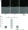
Extracellular ATP-induced chondrocyte cytotoxicity via P2X7 receptors. A Representative bright-field and fluorescence images of active caspase 3/7 signals in chondrocytes. Cells were incubated for 3 h with no treatment (control), ATP (3 mM), A438079 (1 μM), or ATP (3 mM) plus A438079 (1 μM). B Average fluorescence intensity of caspase 3/7 signals obtained with each treatment, normalized to the control values. Bars represent means ± SEM (n = 6). *P < 0.05 vs. control. C ATP-induced LDH release. Cells were incubated for 3 h with no treatment (untreated), A438079 (1 μM), with/without ATP (3 mM). The LDH activity in the medium was quantified by colorimetric assay and normalized to the maximum LDH activity after complete cell lysis to calculate relative LDH release (%). Bars represent means ± SEM (n = 3). *P < 0.05 vs. control
Fig. 8
Fig. 8
ATP-induced PGE2 release via P2X7 activation. Extracellular ATP-induced PGE2 release from articular chondrocytes. Cells were incubated for 1 h with no treatment (untreated), A438079 (1 μM), nominally Ca2+-free medium, ZnCl2 (300 μM), AACOCF3 (10 μM), celecoxib (10 μM), etodolac (10 μM), with/without ATP (3 mM). Cell culture supernatants were collected and PGE2 concentration was measured. The data were normalized to the untreated control value to obtain relative PGE2 release (%). Bars represent means ± SEM (n = 6). *P < 0.05 vs. control, P < 0.05 vs. untreated (ATP)
Fig. 9
Fig. 9
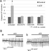
COX-2 inhibitors reduced the P2X7-induced apoptotic signal without current suppression. A Cells were incubated for 3 h with no treatment (untreated), AACOCF3 (10 μM), celecoxib (10 μM), etodolac (10 μM), with/without ATP (3 mM). The fluorescence intensity of caspase-3/7 signals was measured and normalized to the untreated control value. Bars represent means ± SEM (n = 6). *P < 0.05 vs. control. B Effects of celecoxib and etodolac on the ATP-induced current. Celecoxib and etodolac were added to the external solution during ATP (300 μM) exposure. Representative original chart recordings of the membrane current during the experiments are shown

Similar articles

Cited by

References

    1. Burnstock G. Introduction: P2 receptors. Curr Top Med Chem. 2004;4:793–803. doi: 10.2174/1568026043451014. - DOI - PubMed
    1. Khakh BS, North RA. Neuromodulation by extracellular ATP and P2X receptors in the CNS. Neuron. 2012;76:51–69. doi: 10.1016/j.neuron.2012.09.024. - DOI - PMC - PubMed
    1. Le Feuvre R, Brough D, Rothwell N. Extracellular ATP and P2X7 receptors in neurodegeneration. Eur J Pharmacol. 2002;447:261–269. doi: 10.1016/S0014-2999(02)01848-4. - DOI - PubMed
    1. Burnstock G. Purinergic cotransmission. Exp Physiol. 2009;94:20–24. doi: 10.1113/expphysiol.2008.043620. - DOI - PubMed
    1. Garcia M, Knight MM. Cyclic loading opens hemichannels to release ATP as part of a chondrocyte mechanotransduction pathway. J Orthop Res. 2010;28:510–515. - PubMed

Substances

LinkOut - more resources