. 2018 Dec;80(6):2549-2559.
doi: 10.1002/mrm.27236. Epub 2018 May 30.

Revealing sub-voxel motions of brain tissue using phase-based amplified MRI (aMRI)

Affiliations

Revealing sub-voxel motions of brain tissue using phase-based amplified MRI (aMRI)

Itamar Terem et al. Magn Reson Med. 2018 Dec.

Abstract

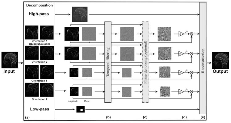
Purpose: Amplified magnetic resonance imaging (aMRI) was recently introduced as a new brain motion detection and visualization method. The original aMRI approach used a video-processing algorithm, Eulerian video magnification (EVM), to amplify cardio-ballistic motion in retrospectively cardiac-gated MRI data. Here, we strive to improve aMRI by incorporating a phase-based motion amplification algorithm.

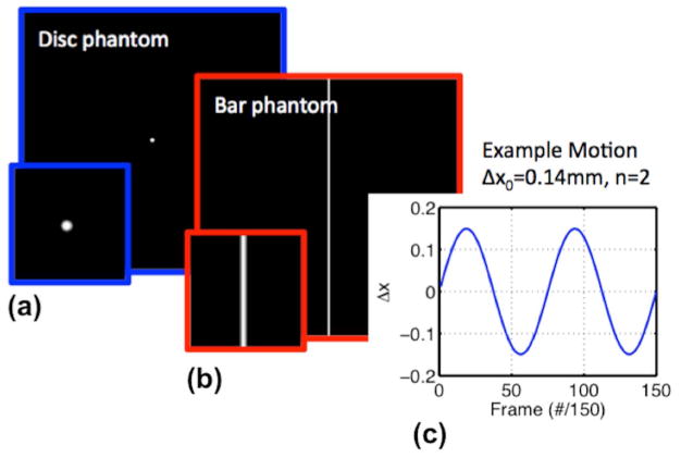
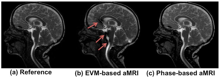
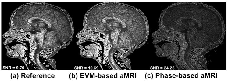
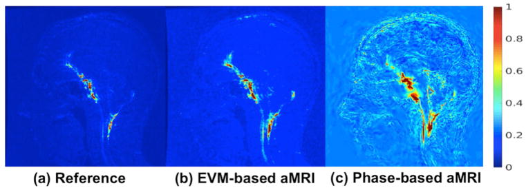
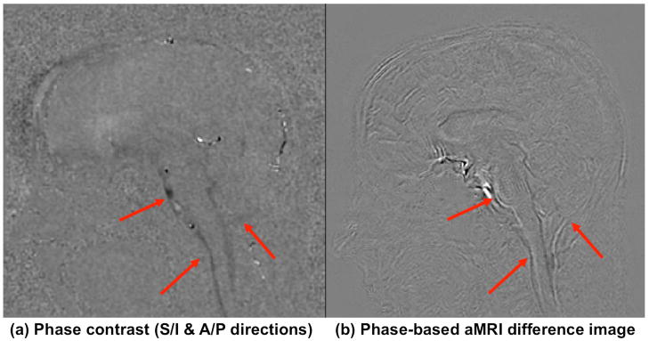
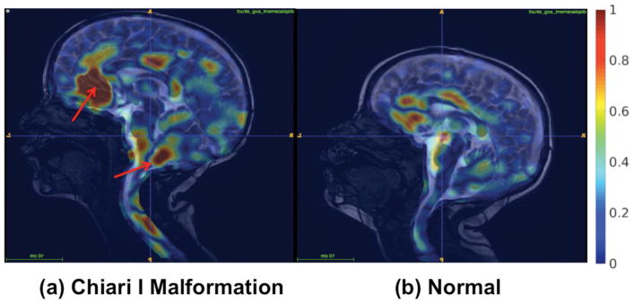
Methods: Phase-based aMRI was developed and tested for correct implementation and ability to amplify sub-voxel motions using digital phantom simulations. The image quality of phase-based aMRI was compared with EVM-based aMRI in healthy volunteers at 3T, and its amplified motion characteristics were compared with phase-contrast MRI. Data were also acquired on a patient with Chiari I malformation, and qualitative displacement maps were produced using free form deformation (FFD) of the aMRI output.

Results: Phantom simulations showed that phase-based aMRI has a linear dependence of amplified displacement on true displacement. Amplification was independent of temporal frequency, varying phantom intensity, Rician noise, and partial volume effect. Phase-based aMRI supported larger amplification factors than EVM-based aMRI and was less sensitive to noise and artifacts. Abnormal biomechanics were seen on FFD maps of the Chiari I malformation patient.

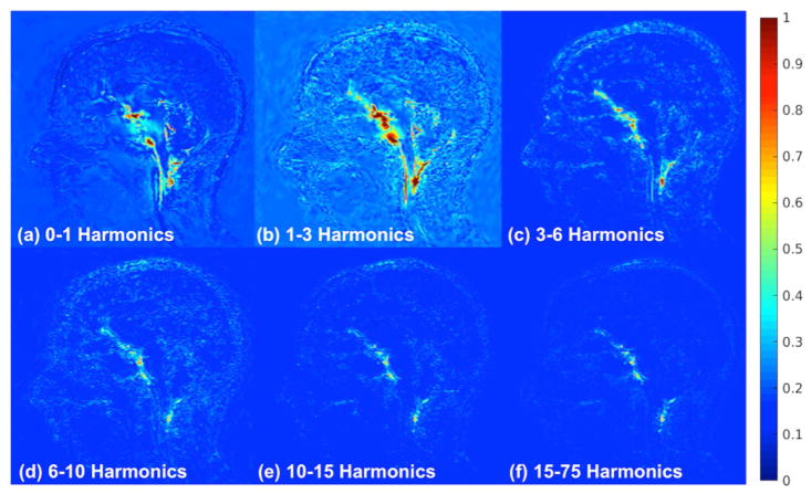
Conclusion: Phase-based aMRI might be used in the future for quantitative analysis of minute changes in brain motion and may reveal subtle physiological variations of the brain as a result of pathology using processing of the fundamental harmonic or by selectively varying temporal harmonics. Preliminary data shows the potential of phase-based aMRI to qualitatively assess abnormal biomechanics in Chiari I malformation.

Keywords: Chiari I malformation; amplified MRI; balanced steady-state free precession (bSSFP); cardiac-gated; free-form deformation; phase contrast MRI; phase-based video motion processing.


Figures

Figure 1
Figure 1
The phase-based motion processing algorithm, described by Wadhwa et al [19], applied to MRI cine data. (a) The cine MRI (short video) is decomposed by the complex steerable pyramid into scales and orientations. (b) The phases are independently temporally filtered at each spatial location, orientation, and scale. (c) Optional step: the filtered phases can be “spatially” filtered again to increase the phase SNR using amplitude-weighted Gaussian spatial smoothing. (d) The filtered phases are multiplied by an amplification parameter and added to the original phase components, and finally the video is reconstructed (e).
Figure 2
Figure 2
The two digital phantoms: (a) A disc (mimicking the cerebellum) with radius r and (b) a vertical bar (mimicking CSF around the midbrain and spinal cord) with width d, were simulated in MATLAB. (c) 1D sinusoidal motion, Δx=Δx0×sin(2πn150t), with amplitude, Δx0, and harmonic number, n.
Figure 3
Figure 3
A snapshot of the cardiac cycle of the original cine MRI data, EVM-based aMRI, and phase-based aMRI. The first harmonic was amplified with an amplification parameter of 6 (other harmonics were attenuated to zero). Arrows denote shading, blooming, and CSF pulsation artifacts seen on EVM-based aMRI.
Figure 4
Figure 4
Noise maps of (a) the original cine images, (b) EVM-based aMRI, and (c) phase-based aMRI. The SNR in the original data and EVM-based aMRI are close in value due to the fact that the EVM algorithm amplifies the motion and the noise all together. On the other hand, the SNR of phase-based aMRI is approximately 2.5 times larger than EVM-based aMRI, since EVM-based aMRI amplifies temporal brightness changes and the amplitude of noise is amplified linearly. In contrast, phase-based modifies phases (not amplitudes), and as such does not increase the magnitude of spatial noise.
Figure 5
Figure 5
Normalized temporal standard deviation of (a) the original (reference) cine MRI data, (b) EVM-based aMRI, and (c) phase-based aMRI. The data was amplified with an amplification parameter of 10 for the temporal harmonics 1–5 (with no spatial filtering). By computing the unamplified cine images, both EVM-based and phase-based aMRI methods magnify near-imperceptible motion near the midbrain, spinal cord, and cerebellum, but phase-based aMRI also reveals subtle motion in the cerebrum.
Figure 6
Figure 6
Normalized temporal variance maps for different harmonic bands as follows: (a) 0–1 (b) 1–3 (c) 3– 6 (d) 6–10 (e) 10–15, and (f) 15–75. The amplification parameter is 10 and the other harmonics are attenuated to zero. The highest amplification achieved can be seen in the 1–3 harmonic band. This was as expected since this band encompasses the natural frequency of the heart beat, however, we can still see that there are contributions by the higher harmonics.
Figure 7
Figure 7
(a) Phase contrast (maximum displacement) and (b) phase-based aMRI (maximum difference from the first frame) maps showing similar motion characteristics in the mid-brain, spinal cord, and cerebellar region (arrows), but with more subtle motion seen in the cortex on phase-based aMRI.
Figure 8
Figure 8
Natural motion of brain structures and spinal cord may be disrupted by pathologies that alter intracranial pressure and/or cerebrospinal fluid (CSF) flow dynamics at the craniocervical junction. Thus, visualization of brain motion may provide clinicians with invaluable information on the nature and extent of the disease. Here, free form deformation (FFD) maps represent the normalized average displacement, and show the potential of the method to differentiate a pediatric patient Chiari I malformation (4yr male) from a normal control (3yr male).

References

    1. Wagshul ME, Eide PK, Maden JR. The pulsating brain: A review of experimental and clinical studies of intracranial pulsatility. Fluids Barriers CNS. 2011;8(1):5–28. - PMC - PubMed
    1. Alperin N, Vikingstad EM, Gomez-Anson B, Levin DN. Hemodynamically independent analysis of cerebrospinal fluid and brain motion observed with dynamic phase contrast MRI. Magn Reson Med. 1996;35:741–754. - PubMed
    1. Chu D, Levin DN, Alperin N. Assessment of the biomechanical state of intracranial tissues by dynamic MRI of cerebrospinal fluid pulsations: A phantom study. Magn Reson Imaging. 1998;16:1043–1048. - PubMed
    1. Hennig J, Wahkloo AK, Koch D, Laubenberger J. Society of Magnetic Resonance in Medicine. Berkeley, California, USA: 1991. The examination of ECG-dependent brain motion with MR-interferography; p. 44.
    1. Feinberg DA, Mark AS. Human brain motion and cerebrospinal fluid circulation demonstrated with MR velocity imaging. Radiology. 1987;163:793–799. - PubMed

Publication types