. 2018 Oct;20(3):294-301.
doi: 10.22074/cellj.2018.5513. Epub 2018 May 15.

Type 1 Diabetes Mellitus: Cellular and Molecular Pathophysiology at A Glance

Affiliations

Type 1 Diabetes Mellitus: Cellular and Molecular Pathophysiology at A Glance

Bahar Saberzadeh-Ardestani et al. Cell J. 2018 Oct.

Abstract

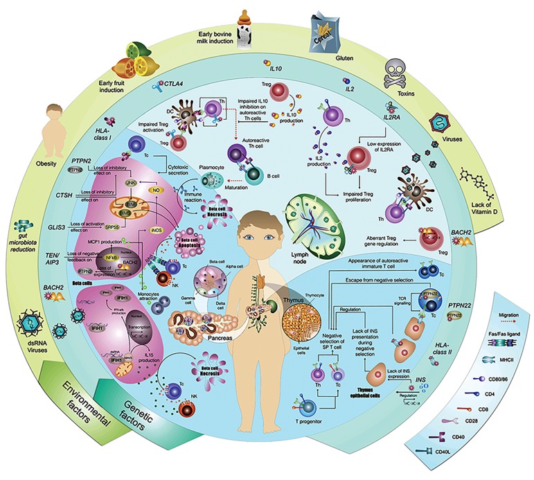
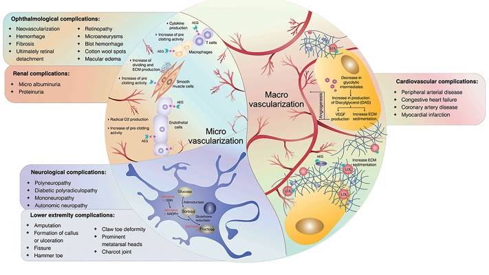
Type 1 diabetes mellitus (T1DM) is a disease where destruction of the insulin producing pancreatic beta-cells leads to increased blood sugar levels. Both genetic and environmental factors play a part in the development of T1DM. Currently, numerous loci are specified to be the responsible genetic factors for T1DM; however, the mechanisms of only a few of these genes are known. Although several environmental factors are presumed responsible for progression of T1DM, to date, most of their mechanisms remain undiscovered. After several years of hyperglycemia, late onsets of macrovascular (e.g., cardiovascular) and microvascular (e.g., neurological, ophthalmological, and renal) complications may occur. This review and accompanying figures provides an overview of the etiological factors for T1DM, its pathogenesis at the cellular level, and attributed complications.

Keywords: Diabetes Complication; Environment; Etiology; Genetic; Type 1 Diabetes Mellitus.


Conflict of interest statement

There is no conflict of interest in this study.

Figures

Fig.1
Fig.1
Genetic, immunologic, and environmental etiologies of type 1 diabetes mellitus (T1DM). The outer circle shows some of the most important environmental etiologies of T1DM and the inner circle presents some of the most important genetic etiologies. The central circle demonstrates each genetic or environmental factor’s known mechanisms of action. The left lower part of the circle shows the dsRNA virus, TEN/AIPS, GLIS3, CTSH, PTPN2 and HLA class 1 mechanism of action at the cellular level in the pancreas microenvironment, which leads to either necrosis or apoptosis of islet beta-cells. The upper part of the circle shows CTLA4, IL10, IL2, IL2RA, BACH2, and viral mechanisms of action in the lymph node. The right lower part of the circle shows PTPN22, HLA class2, and insulin mechanisms of actions which take place in the thymus. AIP3; Actin interacting protein 3, CTLA4; Cytotoxic T-lymphocyte associated protein 4, CTSH; Cathepsin H, GLIS3; GLIS family zinc finger 3, HLA; Human leukocyte antigen, IFIH1; Interferon induced with helicase C domain 1, IL; Interleukin, IL2RA; Interleukin 2 receptor subunit alpha, INS; Insulin, JNK; c-Jun N-terminal kinase, MAVS; Mitochondrial antiviral-signaling, PTPN2; Protein tyrosine phosphatase non-receptor type 2, PTPN22; Protein tyrosine phosphatase non-receptor type 22, BACH2; BTB domain and CNC homolog 2, Tc; Cytotoxic T cell, Th; Helper T cell, NK; Natural killer cell, Treg; Regulatory T cell, DC; Dendritic cell, SP T cell; Single positive T cell, TCR; T cell receptor, and NF-.B; Nuclear factor kappa-light-chain-enhancer of activated B cells.
Fig.2
Fig.2
Chronic complications of type 1 diabetes mellitus (T1DM). T1DM-related chronic complications are divided into two groups based on their pathogenesis: macrovascular and microvascular. The right half of the circle shows the pathogenesis of macrovascular complications [activation of protein C kinase and direct effect of AGEs] and the list of cardiovascular complications. The left half of the circle shows the pathogenesis of microvascular complications (indirect effect of AGEs and defects in polyol metabolism) and the list of related complications. LDL; Low density lipoprotein, AGE; Advanced glycation end product, ECM; Extracellular matrix, VEGF; Vascular endothelial growth factor, GSH; Glutathione, and NADPH; Nicotinamide adenine dinucleotide phosphate.

References

    1. Karvonen M, Pitkaniemi M, Pitkaniemi J, Kohtamaki K, Tajima N, Tuomilehto J. Sex difference in the incidence of insulin-dependent diabetes mellitus: an analysis of the recent epidemiological data.World Health Organization DIAMOND Project Group. Diabetes Metab Rev. 1997;13(4):275–291. - PubMed
    1. Ogurtsova K, da Rocha Fernandes JD, Huang Y, Linnenkamp U, Guariguata L, Cho NH, et al. IDF Diabetes Atlas: Global estimates for the prevalence of diabetes for 2015 and 2040. Diabetes Res Clin Pract. 2017;128:40–50. - PubMed
    1. Patterson CC, Dahlquist GG, Gyurus E, Green A, Soltesz G, Group ES. Incidence trends for childhood type 1 diabetes in Europe during 1989-2003 and predicted new cases 2005-20: a multicentre prospective registration study. Lancet. 2009;373(9680):2027–2033. - PubMed
    1. Steck AK, Rewers MJ. Genetics of type 1 diabetes. Clin Chem. 2011;57(2):176–185. - PMC - PubMed
    1. Knip M, Simell O. Environmental triggers of type 1 diabetes. Cold Spring Harb Perspect Med. 2012;2(7):a007690–a007690. - PMC - PubMed

LinkOut - more resources