. 2018 Oct;57(10):1300-1310.
doi: 10.1002/mc.22844. Epub 2018 Jun 28.

AKR1B10 activates diacylglycerol (DAG) second messenger in breast cancer cells

Affiliations

AKR1B10 activates diacylglycerol (DAG) second messenger in breast cancer cells

Chenfei Huang et al. Mol Carcinog. 2018 Oct.

Abstract

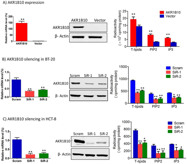
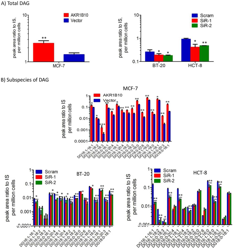
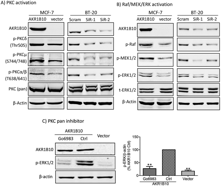
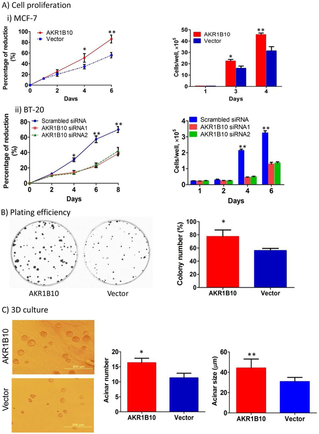
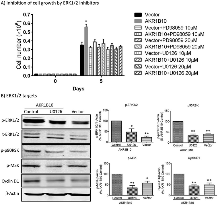
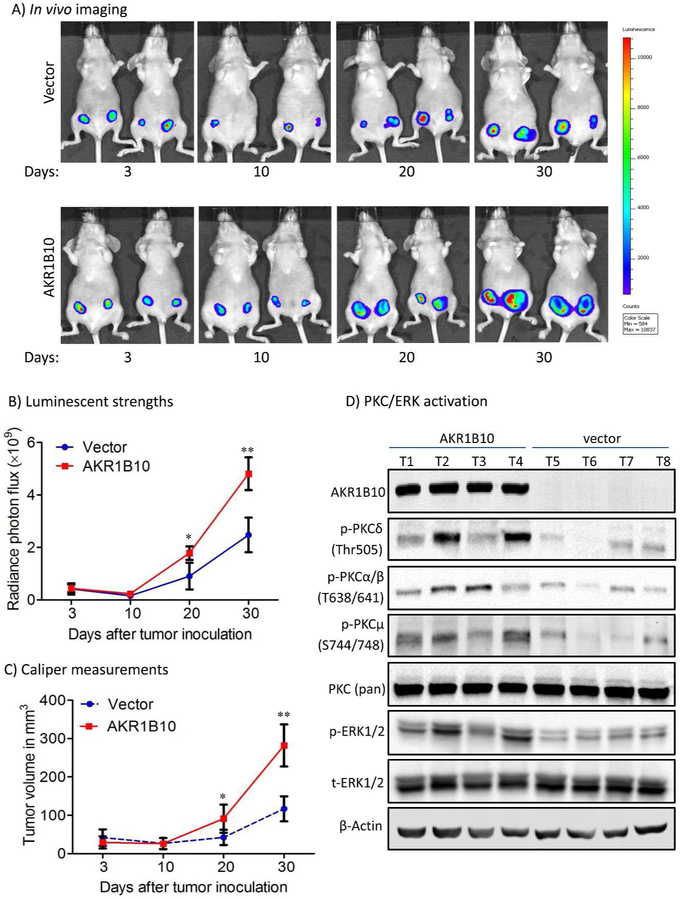
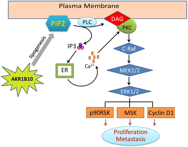
Aldo-keto reductase 1B10 (AKR1B10) is upregulated in breast cancer and promotes tumor growth and metastasis. However, little is known of the molecular mechanisms of action. Herein we report that AKR1B10 activates lipid second messengers to stimulate cell proliferation. Our data showed that ectopic expression of AKR1B10 in breast cancer cells MCF-7 promoted lipogenesis and enhanced levels of lipid second messengers, including phosphatidylinositol bisphosphate (PIP2), diacylglycerol (DAG), and inositol triphosphate (IP3). In contrast, silencing of AKR1B10 in breast cancer cells BT-20 and colon cancer cells HCT-8 led to decrease of these lipid messengers. Qualitative analyses by liquid chromatography-mass spectrum (LC-MS) revealed that AKR1B10 regulated the cellular levels of total DAG and majority of subspecies. This in turn modulated the phosphorylation of protein kinase C (PKC) isoforms PKCδ (Thr505), PKCµ (Ser744/748), and PKCα/βII (Thr638/641) and activity of the PKC-mediated c-Raf/MEK/ERK signaling cascade. A pan inhibitor of PKC (Go6983) blocked ERK1/2 activation by AKR1B10. In these cells, phospho-p90RSK, phospho-MSK, and Cyclin D1 expression was increased by AKR1B10, and pharmacological inhibition of the ERK signaling cascade with MEK1/2 inhibitors U0126 and PD98059 eradicated induction of phospho-p90RSK, phospho-MSK, and Cyclin D1. In breast cancer cells, AKR1B10 promoted the clonogenic growth and proliferation of breast cancer cells in two-dimension (2D) and three-dimension (3D) cultures and tumor growth in immunodeficient female nude mice through activation of the PKC/ERK pathway. These data suggest that AKR1B10 stimulates breast cancer cell growth and proliferation through activation of DAG-mediated PKC/ERK signaling pathway.

Keywords: AKR1B10; PKC/ERK cascade; breast cancer; diacylglycerol; lipid second messengers; phosphatidylinositol bisphosphate.


Conflict of interest statement

Conflict of interest: Authors declare no conflict of interest with the contents of this article.

Figures

Figure 1.
Figure 1.. AKR1B10 promotes lipogenesis of cancer cells.
AKR1B10 expression and lipid synthesis in the MCF-7 (A), BT-20 (B) and HCT-8 (C) cells. Left: AKR1B10 mRNA by qRT-PCR, GAPDH mRNA was used as an internal control; middle: AKR1B10 protein by Western blot; and right: Lipogenesis, showing total lipid (T-lipid), PIP2 and IP3 levels influenced by AKR1B10. Scram, scrambled siRNA; SiR-1, AKR1B10 siRNA-1; and SiR-2, AKR1B10 siRNA-2. *, p<0.05 and **, p<0.01 when compared to vector control.
Figure 2.
Figure 2.. AKR1B10 increases DAG levels in breast cancer cells.
DAG and subspecies were measured quantitatively by liquid chromatography-mass spectrum as described in Methods and Materials. (A) Total DAG levels in MCF-7 cells with AKR1B10 expression (left) and BT-20 cells and HCT-8 cells with AKR1B10 silencing (right). (B) Levels of various subspecies of DAG in MCF-7 (upper), BT-20 (lower left) and HCT-8 (lower right) cells. Y axis is in log10. IS, internal standard. *, p<0.05 and **, p<0.01 when compared to vector control.
Figure 3.
Figure 3.. AKR1B10 activates PKC/ERK signaling pathway in breast cancer cells.
(A) PKC activation by AKR1B10, showing p-PKCδ (Thr505), p-PKCμ (Ser744/748) and p-PKCα/βII (Thr638/641) levels in the MCF-7 with ectopic expression of AKR1B10 and BT-20 cells with silencing of AKR1B10. (B) Raf/MER/ERK activation by AKR1B10, showing p-Raf, p-MEK and p-ERK1/2 levels in the MCF-7 with ectopic expression of AKR1B10 and BT-20 cells with silencing of AKR1B10. (C) ERK inhibition by a broad PKC inhibitor, Go6893 (10μM), showing decreased p-ERK1/2 level by Go6893 in the MCF-7 cells with AKR1B10 expression. Left panel: Quantitative analysis of band intensity. **, p<0.01 when compared to AKR1B10 Control cells. Ctrl: control.
Figure 4.
Figure 4.. AKR1B10 promotes growth and proliferation of breast cancer cells.
(A) Proliferation of MCF-7 and BT-20 cells measured by Alamar blue assays (left) and cell counting assays (right). (B) Plating efficiency of MCF-7 cells in cell culture dishes. (C) Acinar formation and growth of MCF-7 cells in the Matrigel-based 3D culture. *, p<0.05 and **, p<0.01 when compared to vector control.
Figure 5.
Figure 5.. ERK signaling and target genes that are involved in cell proliferation enhanced by AKR1B10.
(A) Inhibition of MCF-7 cell proliferation by MEK inhibitors, PD98059 and U0126 at 10 µM or 20 µM. **, p<0.01 when compared to the vector control or PD98059/U0126-treated cells. (B) ERK targeted genes in MCF-7 cell. The p-ERK1/2, p-p90RSK and p-MSK levels and Cyclin D1 expression were increased by AKR1B10 in the cells, and the increased p-p90RSK, p-MSK and Cyclin D1 levels were eradicated by MEK inhibitor, U0126 (10 µM). Right panel: Quantitative analyses of indicated proteins. *, p<0.05 and **, p<0.01 when compared to AKR1B10 Control cells.
Figure 6.
Figure 6.. AKR1B10 promotes tumor growth in female nude mice.
MCF-7 cells (5 × 10⁶) labeled with luciferase were implanted in the mammary fat pads (orthotopic) of immunodeficient female mice at 4-6 weeks old. A pellet of 17β-estradiol (0.72mg/pellet) was implanted beneath the neck skin. (A) Representative in vivo bioluminescent images at days 3, 10, 20 and 30 post the cell injections. (B) Tumor volumes by in vivo bioluminescence at photon/second. (C) Caliper measurements of the tumor size. *, p<0.05 and **, p<0.01 when compared to vector control. (D) PKC/ERK signaling activity in tumors. *, p<0.05 and **, p<0.01 when compared to vector control.
Figure 7.
Figure 7.. Hypothetic model of cell growth and proliferation promoted by AKR1B10.
In the breast cancer cells, AKR1B10 promotes lipogenesis and increases cellular lipid second messengers PIP2, IP3 and DAG, which activates the PKC/ERK signaling cascade. The p90RSK, MSK and cyclin D1 functions as downstream targets of the ERK1/2 in this process.

References

    1. Santos CR, Schulze A. Lipid metabolism in cancer. The FEBS journal 2012;279(15):2610–2623. - PubMed
    1. Ghaffari P, Mardinoglu A, Nielsen J. Cancer Metabolism: A Modeling Perspective. Frontiers in physiology 2015;6:382. - PMC - PubMed
    1. Spiegel S, Foster D, Kolesnick R. Signal transduction through lipid second messengers. Current opinion in cell biology 1996;8(2):159–167. - PubMed
    1. Currie E, Schulze A, Zechner R, Walther TC, Farese RV Jr. Cellular fatty acid metabolism and cancer. Cell metabolism 2013;18(2):153–161. - PMC - PubMed
    1. Boroughs LK, DeBerardinis RJ. Metabolic pathways promoting cancer cell survival and growth. Nature cell biology 2015;17(4):351–359. - PMC - PubMed

Publication types