. 2018 Jul 30;39(8):1068-1078.
doi: 10.1093/carcin/bgy073.

Intestinal microbiota enhances pancreatic carcinogenesis in preclinical models

Affiliations

Intestinal microbiota enhances pancreatic carcinogenesis in preclinical models

Ryan M Thomas et al. Carcinogenesis. .

Abstract

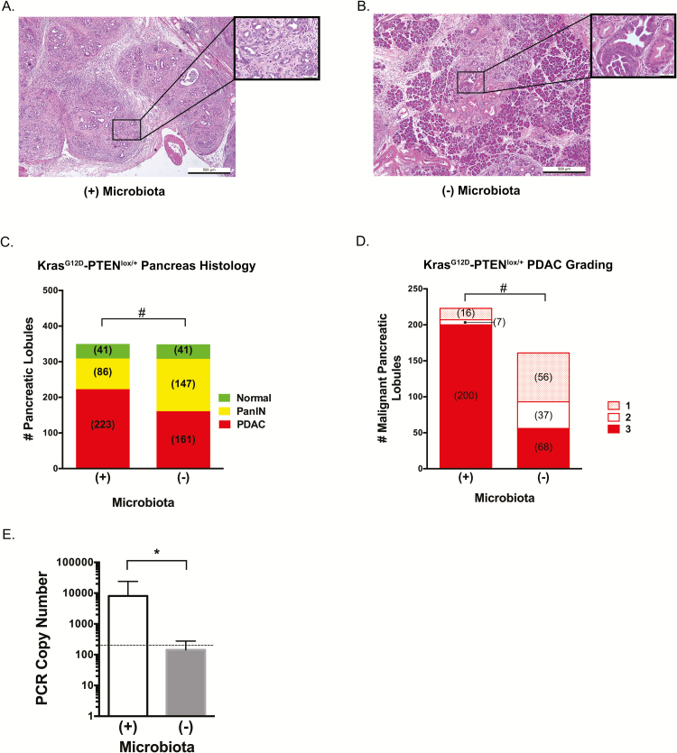
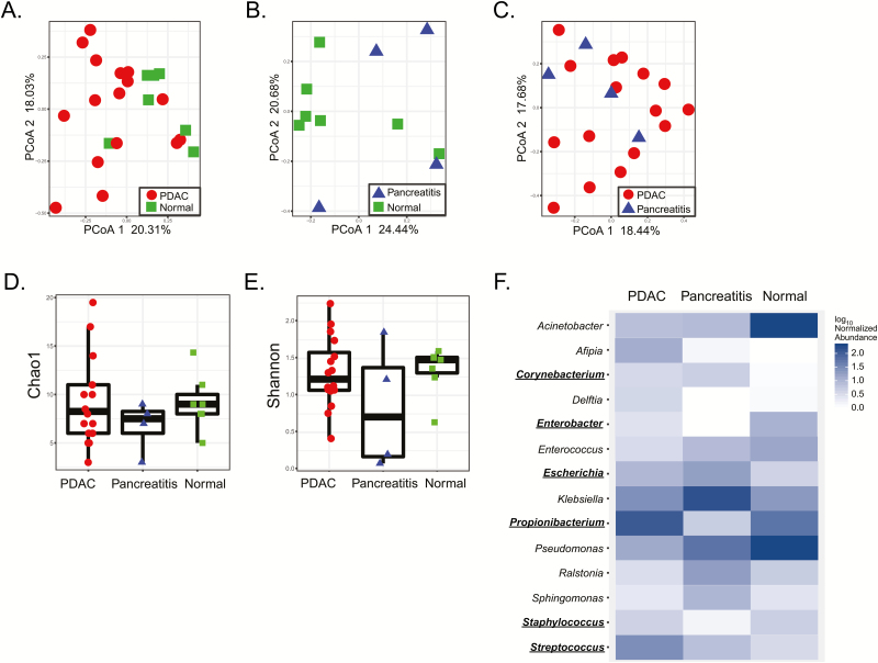
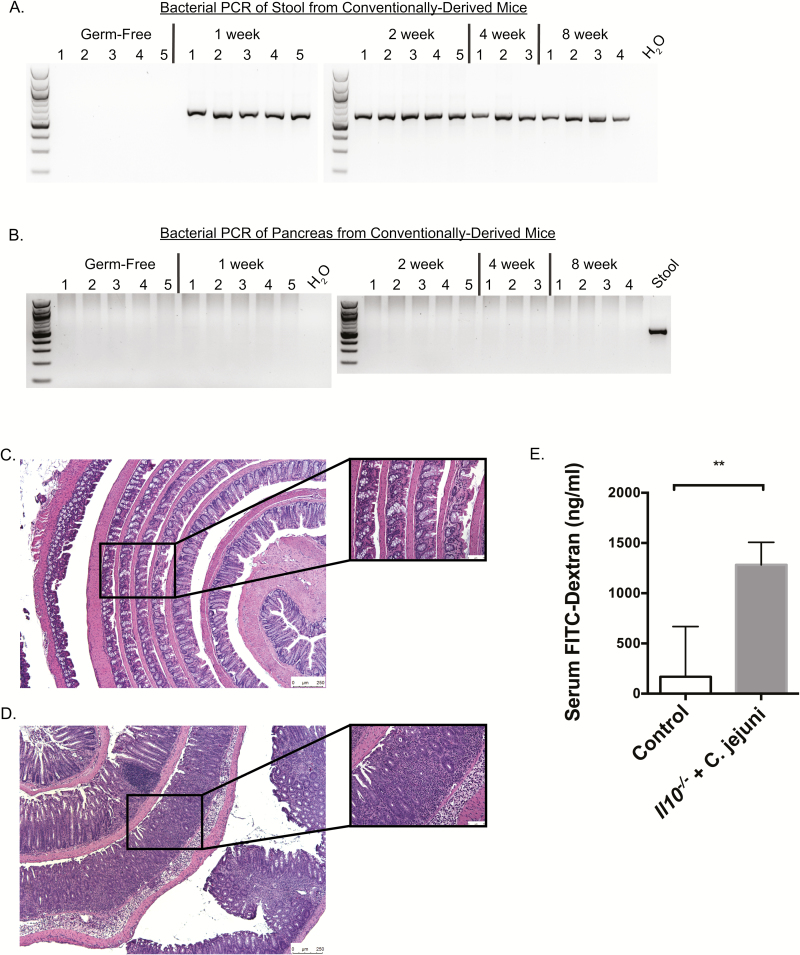
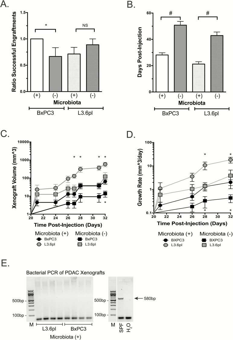
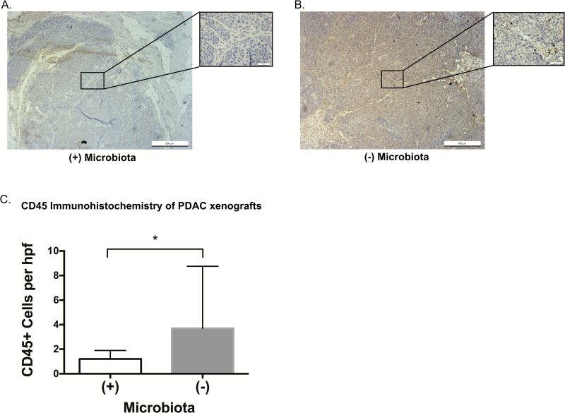
Pancreatic ductal adenocarcinoma (PDAC) is the third leading cause of cancer death in the United States yet data are scant regarding host factors influencing pancreatic carcinogenesis. Increasing evidence support the role of the host microbiota in carcinogenesis but its role in PDAC is not well established. Herein, we report that antibiotic-mediated microbial depletion of KrasG12D/PTENlox/+ mice showed a decreased proportion of poorly differentiated tumors compared to microbiota-intact KrasG12D/PTENlox/+ mice. Subsequent 16S rRNA PCR showed that ~50% of KrasG12D/PTENlox/+ mice with PDAC harbored intrapancreatic bacteria. To determine if a similar observation in humans correlates with presence of PDAC, benign and malignant human pancreatic surgical specimens demonstrated a microbiota by 16S bacterial sequencing and culture confirmation. However, the microbial composition did not differentiate PDAC from non-PDAC tissue. Furthermore, murine pancreas did not naturally acquire a pancreatic microbiota, as germ-free mice transferred to specific pathogen-free housing failed to acquire intrapancreatic bacteria over time, which was not augmented by a murine model of colitis. Finally, antibiotic-mediated microbial depletion of Nod-SCID mice, compared to microbiota-intact, showed increased time to PDAC xenograft formation, smaller tumors, and attenuated growth. Interestingly, both xenograft cohorts were devoid of intratumoral bacteria by 16S rRNA PCR, suggesting that intrapancreatic/intratumoral microbiota is not the sole driver of PDAC acceleration. Xenografts from microbiota-intact mice demonstrated innate immune suppression by immunohistochemistry and differential regulation of oncogenic pathways as determined by RNA sequencing. Our work supports a long-distance role of the intestinal microbiota on PDAC progression and opens new research avenues regarding pancreatic carcinogenesis.


Figures

Figure 1.
Figure 1.
Microbiota accelerates progression to high-grade pancreatic adenocarcinoma in the KrasG12D/PTENlox/+ mouse model. Mixed gender KrasG12D/PTENlox/+ mice were weaned at 1 month of age and the microbiota depleted with broad spectrum antibiotics for an additional 2 months [Microbiota (-)]. Mice were euthanized at 3 months of age and 50 lobules from each harvested pancreas pathologically examined for cancer grade (A, microbiota intact, representative image) and PanIN (B, microbiota depleted, representative image). Images presented at 5× magnification with inset at 40x magnification. The presence of microbiota accelerated the progression of lobules from PanIN to PDAC in the mice with microbiota present (C). Additionally, for KrasG12D/PTENlox/+ mice with cancer, there was a significant difference in PDAC grades between cohorts with grade 3 (poorly differentiated) cancer in the mice with an intact microbiota compared to microbiota depleted (D). Confirmation of microbiota depletion in antibiotic treated mice was made by bacterial PCR using universal primers (E). Dotted line in (E) indicates lower limit of detection for PCR detection, respectively (*P ≤ 0.05; #P < 0.001).
Figure 2.
Figure 2.
The human pancreas harbors a microbiota that does not discriminate between disease states. 16S bacterial DNA sequencing from surgically resected human pancreatic specimens demonstrates no clustering or differences in bacterial populations at the genus level by principle coordinate analysis based on pathologic state of the pancreatic tissue (AC). There was no difference in species richness on Chao1 index (D) or in species diversity on Shannon index (E). Sequencing did, however, demonstrate differences in several bacterial genera between disease states but these were not significant after FDR correction (F). Common genera between 16S sequencing of PDAC tissue and confirmatory 16S sequencing of cultured bacteria from additional independent PDAC tissue samples (Supplementary Table 8, available at Carcinogenesis Online) are underlined in (F).
Figure 3.
Figure 3.
Pancreatic microbes are not acquired from the intestine in a temporal relationship despite altered intestinal barrier function. PCR of stool (A) and pancreas (B) using universal bacterial primers demonstrated the presence of bacteria in stool of conventionally derived mice (germ-free 129SvEv mice that were transferred from gnotobiotic isolators to SPF housing conditions after oral gavage with 1 × 10⁵ SPF bacteria) but failure of the pancreas to acquire microbes after 1, 2, 4 and 8 weeks of intestinal colonization. Electrophoresis blots have been cropped at the top and bottom for the sake of space limitations. Compared to non-gavaged controls (C), the colons of GF IL10/ mice transferred to SPF housing and gavaged with Campylobacter jejuni (D) had increased colitis on haematoxylin and eosin (colitis score: 0.288 versus 3.66, respectively; P < 0.001). Serum FITC-dextran measurement demonstrated increased serum FITC-dextran in C. jejuni gavaged Il10−/− mice compared to non-gavaged controls, confirming increased intestinal permeability (E; **P ≤ 0.01).
Figure 4.
Figure 4.
The intestinal microbiota accelerates pancreatic carcinogenesis in a murine xenograft model. Xenografts were established in 4–6-week-old Nod-SCID mice after the depletion of the intestinal microbiota with broad spectrum antibiotics. After confirmation of microbiota depletion by routine culture methods, the human pancreatic cancer cell lines, BxPC3 and L3.6pl, were heterotopically injected into the flank of the mice. Presence of the intestinal microbiota resulted in improved engraftment efficiency (A), accelerated detection of xenograft growth (B), larger tumors (C) and increased xenograft growth rate (D). Time points of statistical significance for BxPC3 and L3.6pl are located below and above significant values, respectively (*P ≤ 0.05; #P < 0.001). Pancreatic cancer xenografts lack bacterial colonization regardless of antibiotic treatment as isolated bacterial genomic DNA from harvested pancreatic cancer xenografts using a universal 16S bacterial primer failed to detect bacteria (E). Predicted 580bp PCR product from the universal 16S bacterial primer set is indicated (M, molecular weight marker). Electrophoresis blot (E) has been cropped at the top and bottom for the sake of space limitations and controls used from the same PCR reaction but unable to be ran on the same gel are cropped into the right panel of (E).
Figure 5.
Figure 5.
Intact intestinal microbiota associates with decreased CD45 immune cell infiltration in pancreatic cancer xenografts. Immunohistochemistry for CD45 was performed on PDAC xenografts from mice with an intact (A, representative image) or antibiotic depleted (B, rep resentative image) intestinal microbiota. Images presented at 5× magnification with inset at 40× magnification. CD45 positive cells (denoted by arrows) were scored in 10 random high-powered fields (40× ) for each xenograft. There was a statistically significant decrease (C, P = 0.009) in the CD45+ cell infiltration in xenografts derived from microbiota-intact mice (*P ≤ 0.05), suggesting innate immune cell suppression.

References

    1. Hruban R.H., et al. (2000)Progression model for pancreatic cancer. Clin. Cancer Res., 6, 2969–2972. - PubMed
    1. Iacobuzio-Donahue C.A., et al. (2012)Genetic basis of pancreas cancer development and progression: insights from whole-exome and whole-genome sequencing. Clin. Cancer Res., 18, 4257–4265. - PMC - PubMed
    1. Bailey P., et al. ; Pancreatic Cancer Genome Initiative. (2016)Genomic analyses identify molecular subtypes of pancreatic cancer. Nature, 531, 47–52. - PubMed
    1. Brat D.J., et al. (1998)Progression of pancreatic intraductal neoplasias to infiltrating adenocarcinoma of the pancreas. Am. J. Surg. Pathol., 22, 163–169. - PubMed
    1. Hruban R.H., et al. (2001)Pancreatic intraepithelial neoplasia: a new nomenclature and classification system for pancreatic duct lesions. Am. J. Surg. Pathol., 25, 579–586. - PubMed

Publication types

MeSH terms