Case Reports
. 2018 Sep 8;218(8):1249-1260.
doi: 10.1093/infdis/jiy311.

Ultrapotent Human Neutralizing Antibody Repertoires Against Middle East Respiratory Syndrome Coronavirus From a Recovered Patient

Affiliations
Case Reports

Ultrapotent Human Neutralizing Antibody Repertoires Against Middle East Respiratory Syndrome Coronavirus From a Recovered Patient

Peihua Niu et al. J Infect Dis. .

Abstract

Background: The Middle East respiratory syndrome coronavirus (MERS-CoV) causes severe respiratory infection with a high (~35%) mortality rate. Neutralizing antibodies targeting the spike of MERS-CoV have been shown to be a therapeutic option for treatment of lethal disease.

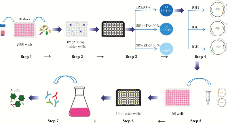
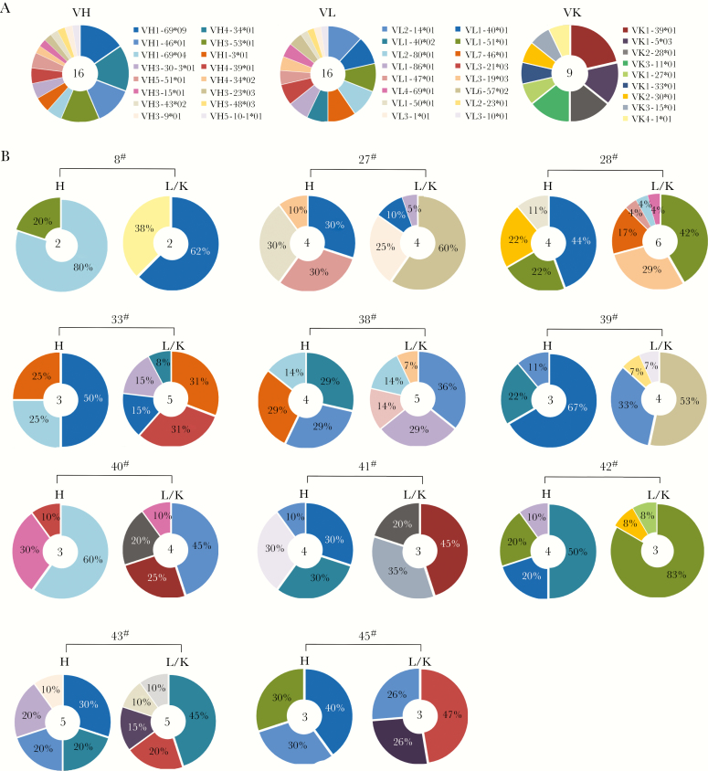
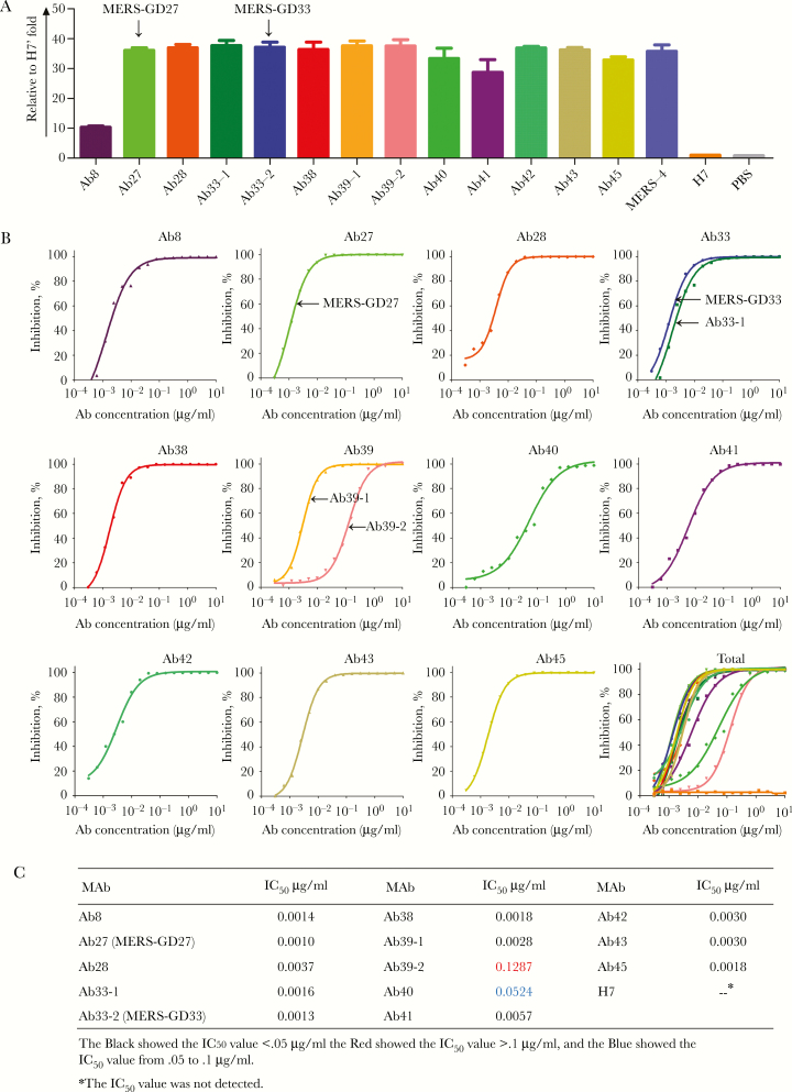
Methods: We describe the germline diversity and neutralizing activity of 13 potent human monoclonal antibodies (mAbs) that target the MERS-CoV spike (S) protein. Biological functions were assessed by live MERS-CoV, pseudotype particle and its variants, and structural basis was also determined by crystallographic analysis.

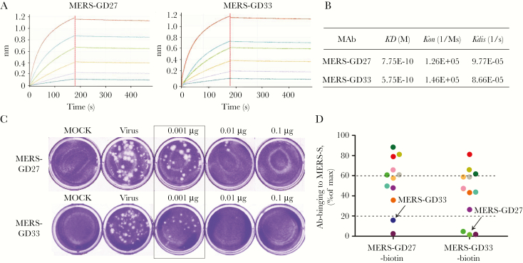
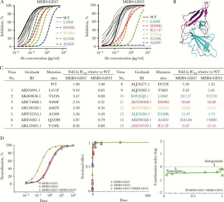
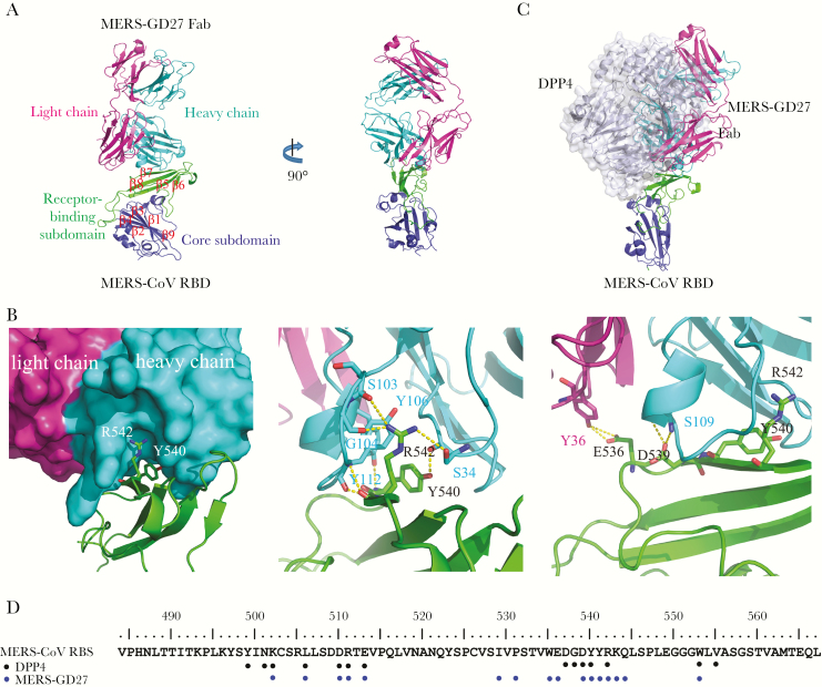
Results: Of the 13 mAbs displaying strong neutralizing activity against MERS-CoV, two with the immunoglobulin heavy-chain variable region (IGHV)1-69-derived heavy chain (named MERS-GD27 and MERS-GD33) showed the most potent neutralizing activity against pseudotyped and live MERS-CoV in vitro. Mutagenesis analysis suggested that MERS-GD27 and MERS-GD33 recognized distinct regions in S glycoproteins, and the combination of 2 mAbs demonstrated a synergistic effect in neutralization against pseudotyped MERS-CoV. The structural basis of MERS-GD27 neutralization and recognition revealed that its epitope almost completely overlapped with the receptor-binding site.

Conclusions: Our data provide new insights into the specific antibody repertoires and the molecular determinants of neutralization during natural MERS-CoV infection in humans. This finding supports additional efforts to design and develop novel therapies to combat MERS-CoV infections in humans.


Figures

Figure 1.
Figure 1.
Workflow for generation of human monoclonal antibodies (mAbs) by cloning antibody genes from primary human B cells. Step 1. B cells were isolated from peripheral blood mononuclear cells of a recovered Middle East respiratory syndrome coronavirus (MERS-CoV) patient and then cultured in the 96-well plates in the presence of 3T3 cells, human interleukin (hIL)-2, CpG2006, and hIL-21 for 10 days. Step 2. Culture supernatants were used to detect MERS-CoV spike (MERS-CoV S) for binding activities using enzyme-linked immunosorbent assay. Step 3. Positive wells were used to detect neutralization activities against MERS-CoV S pseudoviruses. Step 4. The variable regions were cloned into expression vectors and analyzed by sequencing technology. Step 5. The selected VH and VL/VK clonal genes were transiently cotransfected into HEK-293T cells from the same well. Step 6. The culture supernatants were detected against MERS-CoV S pseudoviruses for neutralization activities. Step 7. The neutralizing mAbs were purified, and the immunological function was validated. Abbreviations: IGH, immunoglobulin heavy-chain; IGL, immunoglobulin light chain; IGK, immunoglobulin ; IR, inhibition rate.
Figure 2.
Figure 2.
The germline characteristics of the anti-Middle East respiratory syndrome coronavirus spike monoclonal antibodies (mAbs). (A) Clonal diversity of B cells and (B) the mAbs of 11 wells with exceptionally potent neutralizing activity. The VH, VL, and VK repertoires were shown as a pie chart, with each slice representing a unique VH, VL, and VK clone. The percentage of each slice for A was shown in Supplementary Table S1. The total number of sequences was indicated by the number at the center of each pie chart.
Figure 3.
Figure 3.
Binding activity and neutralizing activity of 13 monoclonal antibodies (mAbs). (A) Binding characterization determined from enzyme-linked immunosorbent assay. Middle East respiratory syndrome coronavirus spike (MERS-CoV S) protein was coated on the 96-well plate 24 hours before the binding test. The mAbs and goat antihuman immunoglobulin (Ig)G Ab/horseradish peroxidase (HRP) antibodies were added sequentially. The absorbance at 450 nm was recorded, and the data presented here was normalized to the value of an irrelevant antibody (H7). The bar chart was depicted by GraphPad Prism 5 software. Data are depicted as the means ± standard deviations from 3 repeats. (B) Neutralization of 13 mAbs against MERS-CoV pseudovirus. Pseudotyped virus was incubated with mAbs before infection of DPP4-expressing Huh-7.5 cells. Luciferase activities were measured, and percentage of neutralization was calculated for 2-fold serial dilutions of each antibody in concentrations from 100 μg/mL to 3 ng/mL. (C) Summary of the half maximal inhibitory concentration (IC50) values of the 13 mAbs.
Figure 4.
Figure 4.
The functional verification of Middle East respiratory syndrome (MERS)-GD27 and MERS-GD33 in vitro. (A) Specific interaction between MERS-CoV S protein and MERS-GD27 and MERS-GD33 characterized by biolayer interferometry. The monoclonal antibodies (mAbs) were captured on the 96-well microplate immobilized with anti-hIgG-Fc and tested for binding with gradient concentrations of MERS-coronavirus spike (CoV S) protein. (B) The apparent dissociation constants were calculated and summarized. (C) Neutralizing activities of MERS-GD27 and MERS-GD33 against live MERS-CoV and against plaque formation of Vero-E6 cells by plaque reduction neutralization test. Three different concentrations of Abs (0.001, 0.01, and 0.1 μg/well) were incubated with 30 plaque-forming units/well live MERS-CoV. Cells were stained with crystal violet at the end of treatment, and the plaques were determined. The inhibitory activity is over 50% in the box. (D) Competition studies among MERS-GD27 and MERS-GD33 with other mAbs. The 96-well plate was first coated with MERS-CoV S 24 hours before the competitive binding test. The mixture of Ab-biotins and other antibodies was incubated in the concentration for 50% of maximal effect. Phosphate-buffered saline was used as a blank control. According to the different competitive binding, 3 groups were created (see Supplementary Table S3).
Figure 5.
Figure 5.
Epitope mapping by mutagenesis of pseudotyped Middle East respiratory syndrome coronavirus (MERS-CoV) and combination effects in neutralizing pseudotyped MERS-CoV for MERS-GD27 and MERS-GD33. (A) Neutralizing analysis of MERS-GD27 and MERS-GD33 against MERS-CoV wild-type (WT) and its variant mutants; site-directed mutagenesis was introduced into the WT receptor-binding domain (RBD) sequence to create 15 mutant RBDs of other strains. Discrepant residues significantly reducing the neutralizing activities were indicated by colored and dashed lines, respectively. (B) The spatial relationship of the critical residues. Six highlighted positions and 9 gray positions on the crystal structure of RBD. (C) Summary of inhibition on infection by MERS-GD27 and MERS-GD33 against all pseudotyped viruses bearing the mutant S glycoprotein relative to WT. (D, left) Percentage of neutralization was calculated for serial 3-fold dilutions of each antibody alone and in combination at constant ratios in a range of concentrations from 81 times to 1/81 of half maximal inhibitory concentrations (IC50s). On the x-axis, a dose of 1 was at the IC50 concentration. (Middle) Fractional effect (FA) plots generated by the CompuSyn program. (Right) Median effect plot of calculated combination index (CI) values (logarithmic) versus FA values, in which a log CI of <0 is synergism and a log CI of >0 is antagonism.
Figure 6.
Figure 6.
The overall structure of Middle East respiratory syndrome coronavirus (MERS-CoV) receptor-binding domain (RBD) in complex with neutralizing antibody MERS-GD27 and the binding interface. (A) A ribbon diagram of the complex in which the RBD core subdomain, RBD receptor binding subdomain, MERS-GD27 heavy chain, and MERS-GD27 light chain are colored blue, green, cyan, and purple, respectively. (B) At the binding interface, the β7 strand of RBD interacts with the MERS-GD27 heavy chain. (C) The structural superimpositions of RBD/MERS-GD27 and RBD/DPP4 complexes. (D) The binding sites of MERS-GD27 and DPP4 on receptor binding subdomain.

References

    1. Zaki AM, van Boheemen S, Bestebroer TM, Osterhaus AD, Fouchier RA. Isolation of a novel coronavirus from a man with pneumonia in Saudi Arabia. N Engl J Med 2012; 367:1814–20. - PubMed
    1. Corti D, Zhao J, Pedotti M, et al. . Prophylactic and postexposure efficacy of a potent human monoclonal antibody against MERS coronavirus. Proc Natl Acad Sci U S A 2015; 112:10473–8. - PMC - PubMed
    1. Wang Y, Liu D, Shi W, et al. . Origin and possible genetic recombination of the Middle East respiratory syndrome coronavirus from the first imported case in china: phylogenetics and coalescence analysis. MBio 2015; 6:e01280–15. - PMC - PubMed
    1. Memish ZA, Mishra N, Olival KJ, et al. . Middle East respiratory syndrome coronavirus in bats, Saudi Arabia. Emerg Infect Dis 2013; 19:1819–23. - PMC - PubMed
    1. Ithete NL, Stoffberg S, Corman VM, et al. . Close relative of human Middle East respiratory syndrome coronavirus in bat, South Africa. Emerg Infect Dis 2013; 19:1697–9. - PMC - PubMed

Publication types

MeSH terms