. 2018 Jul 27;46(13):6627-6641.
doi: 10.1093/nar/gky451.

The archaeal ATPase PINA interacts with the helicase Hjm via its carboxyl terminal KH domain remodeling and processing replication fork and Holliday junction

Affiliations

The archaeal ATPase PINA interacts with the helicase Hjm via its carboxyl terminal KH domain remodeling and processing replication fork and Holliday junction

Binyuan Zhai et al. Nucleic Acids Res. .

Abstract

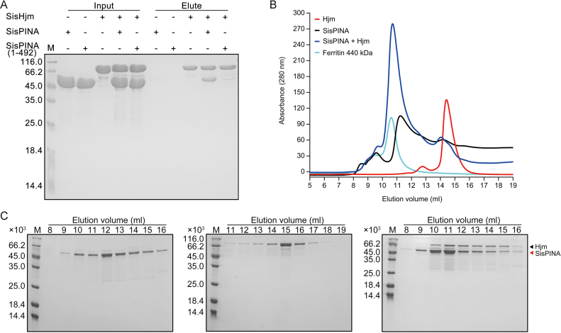
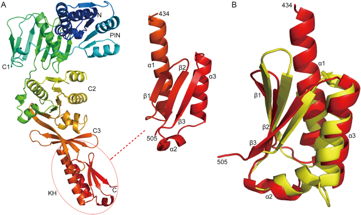
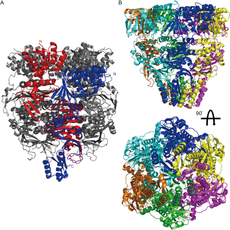
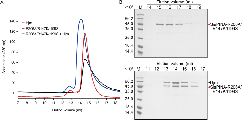
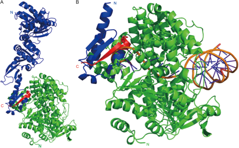
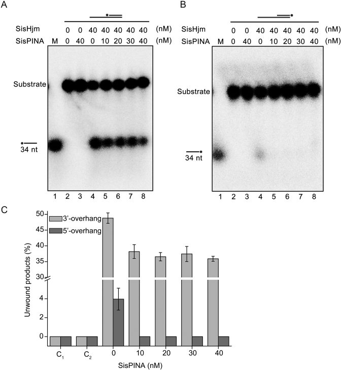
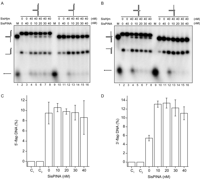
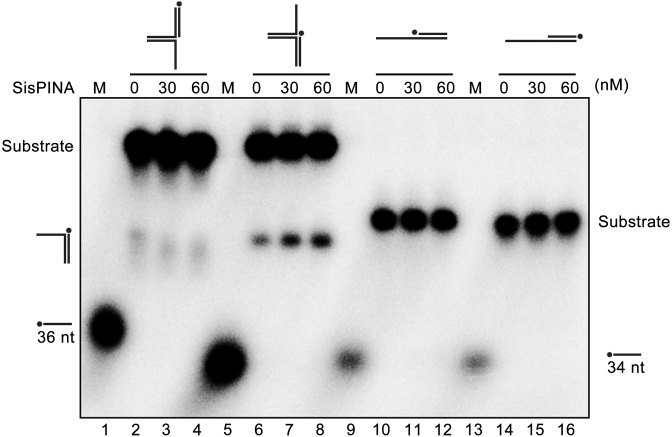
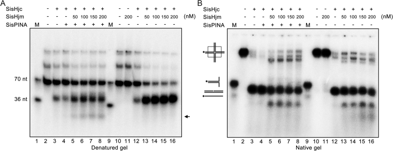
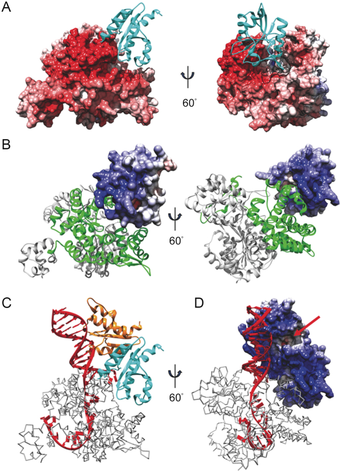
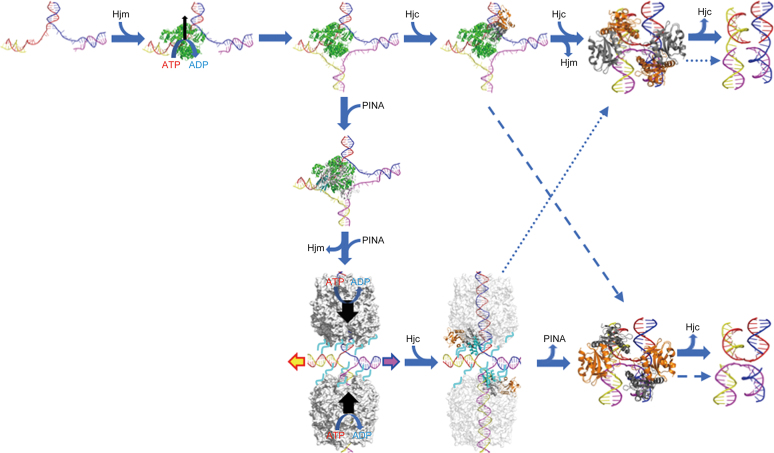
PINA is a novel ATPase and DNA helicase highly conserved in Archaea, the third domain of life. The PINA from Sulfolobus islandicus (SisPINA) forms a hexameric ring in crystal and solution. The protein is able to promote Holliday junction (HJ) migration and physically and functionally interacts with Hjc, the HJ specific endonuclease. Here, we show that SisPINA has direct physical interaction with Hjm (Hel308a), a helicase presumably targeting replication forks. In vitro biochemical analysis revealed that Hjm, Hjc, and SisPINA are able to coordinate HJ migration and cleavage in a concerted way. Deletion of the carboxyl 13 amino acid residues impaired the interaction between SisPINA and Hjm. Crystal structure analysis showed that the carboxyl 70 amino acid residues fold into a type II KH domain which, in other proteins, functions in binding RNA or ssDNA. The KH domain not only mediates the interactions of PINA with Hjm and Hjc but also regulates the hexameric assembly of PINA. Our results collectively suggest that SisPINA, Hjm and Hjc work together to function in replication fork regression, HJ formation and HJ cleavage.


Figures

Figure 1.
Figure 1.
SisPINA has physical interaction with SisHjm. Analysis of the interaction between SisPINA and SisHjm by pull-down and gel filtration. (A) Recombinant SisPINA or SisPINA (1–492) (without His-tag) was incubated with N-terminal His-tagged SisHjm. Inputs and elution fractions were analyzed by SDS-PAGE with Coomassie blue staining. M, molecular size markers. ‘–’, no protein, ‘+’, protein added. Molecular masses of standard proteins are indicated at the left of each panel. (B) Gel filtration profile of Hjm (red), SisPINA (black), Hjm/SisPINA mixture (blue), and protein marker ferritin (cyan, 440 kD) (GE Healthcare, UK). The protein sample (500 ul) of SisPINA (430 μg), Hjm (230 μg), or their mixture was loaded onto the Superdex 200 column which was equilibrated with the buffer containing 25 mM Tris–HCl, pH 8.0, 200 mM NaCl, and 5% glycerol. (C) SDS-PAGE analysis of the elute fractions in (B). The fractions (1 ml each) were collected and analyzed.
Figure 2.
Figure 2.
The C-terminus of SisPINA folds into a KH domain. (A) The structure of SisPINA-R206A/R147K/I199S. The C-terminal domain (dashed circle) was enlarged and the α-helices and β-strands are indicated. (B) Alignment of SisPINA C-terminal KH domain with NusA. The ribbon structure of the C-terminus of SisPINA (residues 434–505) (red) is superimposed on the structure of the NusA (yellow) from Aeropyrum pernix (PDB ID: 2CXC).
Figure 3.
Figure 3.
Hexameric assembly destabilizes the KH domain of SisPINA. (A) Alignment of the SisPINA-R206A/R147K/I199S structure (blue) with one subunit (red) of the SisPINA-R206A hexamer structure (PDB ID: 5F4H; gray) over the ATPase domain. (B) KH domains clashing among subunits of the hexameric PINA. Six SisPINA-R206A/R147K/I199S monomers (in different colors) are aligned to the SisPINA-R206A hexamer (not shown) over the ATPase domains.
Figure 4.
Figure 4.
Analysis of the physical interaction between SisPINA-R206A/R147K/I199S and SisHjm by gel filtration. (A) Gel filtration profile of Hjm (red), SisPINA-R206A/R147K/I199S (black), and the mixture of Hjm and SisPINA-R206A/R147K/I199S (blue). (B) The SDS-PAGE of eluted fractions of SisPINA-R206A/R147KI/199S (top) and its mixture with Hjm (bottom). The procedure for the analysis was same as that for the analysis of the interaction between SisPINA and Hjm.
Figure 5.
Figure 5.
Complex model of SisPINA interacting with Hjm. (A) Docking result showing that SisPINA (blue) interacts with Hjm (green) through its C-terminal residues 492–502 (red). (B) SisPINA KH domain (colored as in A) clashing with the ssDNA (orange) bound with Hjm (green). DNA positioned via alignment of PfuHjm:DNA complex (PDB ID: 2P6R) with Hjm.
Figure 6.
Figure 6.
Effect of SisPINA on the unwinding activity of SisHjm towards 3′-overhang (84 mer/34 mer) (A) and 5′-overhang (84 mer/34 mer) (B). The ‘*’ indicates labeling of the substrates at the 5′ end. Various concentrations of SisPINA (0, 10, 20, 30 and 40 nM, as hexamer) and 40 nM SisHjm (as monomer) were added into the reaction mixture and the reactions were carried out at 45°C for 25 min. The products were separated on 8% native PAGE gels and analyzed using a phosphor imager. (C) Quantification of the results in (A) and (B). The data were obtained with ImageJ software (NIH) and the values were calculated from at least three repeats.
Figure 7.
Figure 7.
SisPINA and SisHjm in combination remodel the replication fork. SisPINA and SisHjm were mixed to detect the capability of replication fork DNA processing. The fork substrates were labeled at the leading strand (A) or lagging strand (B) and the representative gels are shown. All the reactions were carried out at 45°C for 30 min. The ‘*’ indicates labeling of the substrates at the 5′ end. (C) Effect of SisPINA on the generation of 5′-flap DNA from a pseudo replication fork by Hjm. (D) Effect of SisPINA on the generation of 3′-flap DNA from a pseudo replication fork by Hjm. The products of 5′-flap DNA (A, lanes 2–8) and 3′-flap DNA (B, lanes 2–8) were quantified by Image J (NIH). The values were based on data of at least three experimental repeats.
Figure 8.
Figure 8.
SisPINA is able to unwind 3′-flap DNA. Lanes 2–4, replication fork without lagging strand; lanes 6–8, replication fork without leading strand; lanes 10–12, 3′-overhang DNA; lanes 14–16, 5′-overhang DNA. Lanes 1 and 5, 32P-labeled 36 nt ssDNA marker; Lanes 9 and 13, 32P-labeled 34 nt ssDNA markers. The ‘*’ indicates labeling of the substrates at the 5′ end. The concentrations of SisPINA (as hexamer) used for the analysis were indicated. All of the reactions were performed at 45°C for 30 min and the products were separated on 8% native page gels and analyzed with a phosphor imager.
Figure 9.
Figure 9.
Holliday junction processing by SisPINA, SisHjm, and SisHjc. The cleavage of HSL (mobile) HJ substrate by SisHjc in the presence of SisPINA and SisHjm at indicated concentrations was analyzed by denatured (A) and native (B) gel electrophoresis. All of the reactions were carried out at 55°C for 30 min. The black arrow indicates band that is produced by HJ branch migration and cleavage. The ‘*’ indicates labeling of the substrates at the 5′ end.
Figure 10.
Figure 10.
Modeling Hjm:Hjc unwinding and cleavage of DNA. Docking result showing (A) the electrostatic surface of Hjm interacting with Hjc (cyan ribbons), or (B) the electrostatic surface of Hjc interacting with Hjm (ribbons). The Hjc-interaction region of Hjm is highlighted by green. Model of a Hjc dimer (cyan and orange ribbons (C) or electrostatic surface (D)) bound with Hjm (gray trace) and DNA (red) from PfuHjm:DNA complex (PDB ID: 2P6R) based on results in A&B. The Hjc active site is indicated with a red arrow. Red surface - negative charge, white surface - neutral, blue surface - positive charge.
Figure 11.
Figure 11.
A proposed model for PINA/Hjm/Hjc to process a stalled DNA replication fork. A stalled DNA replication fork (blue, red, yellow, and magenta strands) is recognized and bound by Hjm (green), which proceeds to regress the replication fork and form a HJ structure. Either Hjc (right) or PINA (down) is recruited to the HJ structure by Hjm. The Hjc dimer (gray and orange) binding to the HJ facilitates DNA handover from Hjm to Hjc, prompting Hjm dismissal and turning the open HJ into an X-HJ structure that is nicked by two Hjc dimers (PDB: 2WJ0). Alternatively, a PINA (white) monomer is recruited by Hjm through the interaction of Hjm and the PINA KH domain (cyan), leading to the assembly of the PINA hexamer on the HJ DNA and the dissociation of Hjm. Two PINA hexamers (white surface) are formed on two opposing HJ arms and mediate HJ migration powered by ATP hydrolysis. Two Hjc dimers then bind to the HJ and interact with the PINA C-terminal domains, destabilizing the PINA hexamers. After PINA hexamers are released, the two Hjc dimers turn the open HJ into the X-HJ structure and mediate two symmetric cuts. HJ migration directionality is indicated by colored arrows. Preferred HJ cleavages by Hjc are indicated with solid arrows while alternative HJ cleavages are indicated with dashed arrows. Unfolded PINA KH domains are indicated by cyan lines.

Similar articles

Cited by

References

    1. Branzei D., Foiani M.. Maintaining genome stability at the replication fork. Nat. Rev. 2010; 11:208–219. - PubMed
    1. Kitao H., Iimori M., Kataoka Y., Wakasa T., Tokunaga E., Saeki H., Oki E., Maehara Y.. DNA replication stress and cancer chemotherapy. Cancer Sci. 2018; 109:264–271. - PMC - PubMed
    1. Barlow J.H., Nussenzweig A.. Replication initiation and genome instability: a crossroads for DNA and RNA synthesis. Cell. Mol. Life. Sci. 2014; 71:4545–4559. - PMC - PubMed
    1. Friedberg E.C., Walker G.C., Siede W.. DNA Repair and Mutagenesis. 2005; Washington, D.C: ASM Press.
    1. Atkinson J., McGlynn P.. Replication fork reversal and the maintenance of genome stability. Nucleic Acids Res. 2009; 37:3475–3492. - PMC - PubMed

Publication types

MeSH terms