Review
. 2018 Jun 27;118(12):5952-5984.
doi: 10.1021/acs.chemrev.8b00277. Epub 2018 May 30.

Cell-Wall Recycling of the Gram-Negative Bacteria and the Nexus to Antibiotic Resistance

Affiliations
Review

Cell-Wall Recycling of the Gram-Negative Bacteria and the Nexus to Antibiotic Resistance

David A Dik et al. Chem Rev. .

Abstract

The importance of the cell wall to the viability of the bacterium is underscored by the breadth of antibiotic structures that act by blocking key enzymes that are tasked with cell-wall creation, preservation, and regulation. The interplay between cell-wall integrity, and the summoning forth of resistance mechanisms to deactivate cell-wall-targeting antibiotics, involves exquisite orchestration among cell-wall synthesis and remodeling and the detection of and response to the antibiotics through modulation of gene regulation by specific effectors. Given the profound importance of antibiotics to the practice of medicine, the assertion that understanding this interplay is among the most fundamentally important questions in bacterial physiology is credible. The enigmatic regulation of the expression of the AmpC β-lactamase, a clinically significant and highly regulated resistance response of certain Gram-negative bacteria to the β-lactam antibiotics, is the exemplar of this challenge. This review gives a current perspective to this compelling, and still not fully solved, 35-year enigma.


Conflict of interest statement

The authors declare no competing financial interest.

Figures

Figure 1.
Figure 1.
A comparison of Gram-negative and Gram-positive cell-wall flux. The events of cell-wall biosynthesis, recycling, and turnover are depicted. In rod-shaped Gram-negative bacteria, the PG of the cell wall is found within the periplasm. It has perhaps a single monolayer thickness in its sidewall, and a bi or trilayer thickness at the septa. In contrast, the Gram-positive PG is a multilayered exoskeleton. Together, de novo cell wall and recycled cell wall are manufactured in the cytoplasm and in final form as lipid II are translocated to the periplasm, where they experience a diverse array of structural modification to accommodate many biological pathways. In both types of bacteria, although less frequent in Gram-positive bacteria, the cell wall is recycled. Adapted with permission from ref . Copyright 2013 New York Academy of Sciences.
Figure 2.
Figure 2.
β-Lactam antibiotics mimic the native acyl-d-alanine-d-alanine substrate of the PBP enzymes. The β-lactam ring of the penicillin (left) and the central amide bond of the d-Ala-d-Ala peptide (right) are shown in red.
Figure 3.
Figure 3.
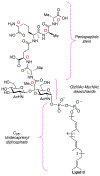
Structure of lipid II (undecaprenyl diphosphate-MurNAc-pentapeptide-GlcNAc) of the Gram-negative bacterium. The stereochemistry of the lactyl moiety of the MurNAc saccharide, and of each of the stereogenic carbons of the amino acids of the peptide stem of the MurNAc (l or d), is shown by the red-colored labels.
Figure 4.
Figure 4.
(a) The divisome protein complex forms at the midcell and facilitates septation. The divisome tracks along the midcell (see green line). (b) The elongasome protein complex forms at the lateral sidewalls and facilitates sidewall expansion. The elongasome tracks along the lateral sidewall (see blue line). Notably absent from modern interpretations of these systems are the lytic transglycosylases, whose participation, although certain, remains undefined. Adapted with permission from ref . Copyright 2016 eLife Sciences Publications (https://creativecommons.org/licenses/by/4.0/).
Figure 5.
Figure 5.
(a) PBPs elongate nascent PG (TG, transglycosylase domain) and incorporate it into the PG macromolecule (TP, transpeptidase domain). (b) β-Lactam antibiotics inhibit the incorporation of nascent PG (GlcNAc, yellow hexagon; MurNAc, green hexagon; anhMurNAc, red hexagon) into the cell wall, resulting in an aberrant form shown. (c–e) The aberrant PG might be misincorporated, resulting in the inevitable death of the bacterium. Slt degrades the nascent aberrant PG in an attempt at onset of repair to protect the bacterium. Adapted with permission from ref . Copyright 2018 United States National Academy of Sciences.
Figure 6.
Figure 6.
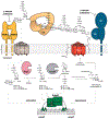
β-Lactam antibiotics inhibit the transpeptidase (TP) domain of the PBPs. The unhindered transglycosylase (TG) domain of bifunctional HMM PBPs continues the lengthening of the nascent PG chain. Nascent PG retains a pentapeptide stem. Slt cleaves the accumulated nascent PG as an effort toward repair. Slt-liberated PG is transported to the cytoplasm by the AmpG permease, where in current form as GlcNAc-1,6-anhMurNAc-pentapeptide (1c) it activates AmpR. NagZ recognizes compound 1c as a substrate and catalyzes the formation of 1,6-anhMurNAc (2c), which also functions to activate AmpR. Both AmpR activators are substrates for AmpD, which catalyzes the formation of recycling intermediates GlcNAc and free pentapeptide (3c). GlcNAc and 3c are recycled by the bacterium in an effort to make new cell wall. Cell-wall precursor UDP-MurNAc-pentapeptides (4) is the AmpR repressor and exists in both homeostasis and β-lactam challenge. The increased concentration of 1c or 2c sufficiently displaces homeostatic concentration of 4 during β-lactam challenge, triggering AmpR activation and β-lactamase transcription.
Figure 7.
Figure 7.
Architecture of LTTR oligomers, as revealed in X-ray structure determinations, adopts distinct conformations, which are defined by a three-scheme classification. Each full-length LTTR comprises a DNA-binding domain, a region of difference I effector-binding domain subdomain (I), and a region of difference II effectorbinding domain subdomain (II). (a) Scheme I identifies LTTRs with weak interactions of the α10–α10 (yellow highlight) region as a result of an offset arrangement of the effector-binding domains. (b) Scheme II identifies LTTRs with separated α10–α10 regions. (c) Scheme III identifies LTTRs with strong interactions of the α10–α10 region (yellow highlight) as a result of extensive surface interactions of the effector-binding domains. Reproduced with permission from ref . Copyright 2008 Microbiology Society (https://creativecommons.org/licenses/by/3.0/).
Figure 8.
Figure 8.
Full-length structure of DNTR was solved by small-angle X-ray scattering (SAX) and a model was proposed by which DNTR could bind its requisite DNA In a repressed compact conformation, DNTR binds to the RBS and ABS′. In an activated extended conformation, DNTR binds RBS and ABS″. (a) The model of DNTR is depicted, whereby the requisite DNA bends at a 240° angle in the repressed conformation and is relaxed to a 94° angle in the active conformation. Importantly it has been surmised that not all LTTRs bend DNA to the same degree. (b) A second model is shown that depicts how an LTTR could bind to its requisite DNA, if the DNA was bent to a lesser degree. In this model, the repressed conformation bends DNA at a 100° angle, and the active conformation bends DNA at a 50° angle. This model stipulates that the dimers of the LTTR tetramer cannot interact in the repressed conformation. Reproduced with permission from ref . (https://creativecommons.org/licenses/by/4.0/). Image courtesy of Prof. Gordon A. Leonard.
Scheme 1.
Scheme 1.. PBP Inactivation by β-Lactam Acylation of the Catalytic Serine Residue of the PBPa
a Although the resulting acyl-enzyme eventually could undergo hydrolysis, restoring the active PBP, the time scale for this hydrolysis well exceeds that of the viability of the bacterium.
Scheme 2.
Scheme 2.
Catalytic Deactivation of the β-Lactam Antibiotic (Here Represented As a Penicillin) by β-Lactamase (Here Represented As a Class C Serine-Dependent Enzyme) by Hydrolytic Opening of Its β-Lactam Ring.
Scheme 3.
Scheme 3.. Turnover of the Gram-Negative Cell-Wall Muropeptides Is Showna
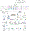
aThe disaccharide is disassembled by a NagZ glucosaminidase, and the peptide stem is separated by the AmpD amidase. Turnover of cell-wall saccharides is an independent pathway from that of peptide turnover. The resulting saccharide pool, coupled with de novo synthesized saccharides, undergo biosynthesis culminating in the cell-wall precursor lipid II. Notably, the lipid II precursor UDP-MurNAc pentapeptide (4) and the cell-wall recycled muropeptides GlcNAc-1,6-anhMurNAc pentapeptide (1c) and 1,6-anhMurNAc pentapeptide (2c) serve as important effectors in the regulation of antibiotic resistance.
Scheme 4.
Scheme 4.
Biosynthetic Transformations (MurA–MurF) in the Mur Pathway Leading from UDP-GlcNAc to UDP-MurNAc-pentapeptide (4)
Scheme 5.
Scheme 5.. Biosynthesis, Modifications, and Degradation of the Gram-Negative Cell-Wall Macromoleculea
aPBP glycosyltransferases assemble Lipid II by accretion. The nascent PG chain exists in pentapeptide form. PBP carboxypeptidases cleave the terminal d-Ala of the peptide stem in an effort to regulate the degree of cell-wall crosslinking. PBP transpeptidases crosslink nascent PG to the cell-wall macromolecule in a process that allows for selective incorporation of new PG at specific sites. PBPs and amidases modify the PG to accommodate various biological events including pili or flagellum formation, secretion systems assembly, elongation, and division. The lytic transglycosylases cleave PG in a unique reaction that forms a 1,6-anhydromuropeptide. These muropeptides are substrates for the AmpG permease, which transports muropeptides to the cytoplasm for recycling.
Scheme 6.
Scheme 6.. Glycosyltransferase Reaction Whereby Nascent PG Chain Is Formed by Catalysis of Adjacent Lipid II Moleculesa
aBifunctional HMM PBPs, RodA, and FtsW perform this reaction, which subsequently releases the lipid II acceptor strand from the membrane. The chemistry likely involves the formation of a reactive oxocarbenium species at the anomeric carbon of MurNAc (not depicted), as either an intermediate or transition state.
Scheme 7.
Scheme 7.. Crosslinking of the Cell-Wall Macromolecule Is an Essential Step in Cell-Wall Biosynthesisaa
aInhibition of this process, by the β-lactam antibiotic, leads to lysis of the bacterium. The crosslinking reaction shown is a 4,3-crosslink, although 3,3-crosslinks also form. Monofunctional and bifunctional HMM PBPs crosslink the cell-wall polymer.
Scheme 8.
Scheme 8.. Amidases Catalyze Hydrolysis of the Stem Peptide of the Muropeptides, and Produce Free Peptide and Denuded (or “Naked”) Glycan As Productsa
aDenuded cell wall in the periplasm at the septum is an important component of cell division. While in the cytoplasm, amide hydrolysis is an essential step in muropeptide turnover.
Scheme 9.
Scheme 9.. LTs Catalyze a Unique Non-Hydrolytic Transacetalization Reaction That Cleaves the MurNAc-GlcNAc β-(1,4)-Glycosidic Bond and Converts MurNAc to 1,6-anhMurNAca
aThe products of LT reactions are specific substrates for the AmpG permease, which allows for cytoplasmic transport and subsequent recycling. The formation of the discrete oxazolinium intermediate might not be relevant for all LTs.
Scheme 10.
Scheme 10.. Turnover of GlcNAc-anhMurNAc Involves an Aspartic Acid Covalent Intermediate, Two Transition State Species Each Invoking an Oxocarbenium, and a Dramatic Conformational Protein Rearrangementa
a The reaction results in the hydrolysis of the GlcNAc-MurNAc β-(1,4)-glycosidic bond.

References

    1. Höltje JV Growth of the Stress-bearing and Shape-maintaining Murein Sacculus of Escherichia coli. Microbiol. Mol. Biol. Rev 1998, 62, 181–203. - PMC - PubMed
    1. Zhao H; Patel V; Helmann JD; Dörr T Don’t Let Sleeping Dogmas lie: New Views of Peptidoglycan Synthesis and its Regulation. Mol. Microbiol 2017, 106, 847–860. - PMC - PubMed
    1. van der Ploeg R; Verheul J; Vischer NOE; Alexeeva S; Hoogendoorn E; Postma M; Banzhaf M; Vollmer W; den Blaauwen T Colocalization and Interaction Between Elongasome and Divisome During a Preparative Cell Division Phase in Escherichia coli. Mol. Microbiol 2013, 87, 1074–1087. - PubMed
    1. Labischinski H; Goodell EW; Goodell A; Hochberg ML Direct Proof of a “More-than-single-layered” Peptidoglycan Architecture of Escherichia coli W7: A Neutron Small-angle Sattering Study. J. Bacteriol 1991, 173, 751–756. - PMC - PubMed
    1. Johnson JW; Fisher JF; Mobashery S Bacterial Cell-Wall Recycling. Ann. N. Y. Acad. Sci 2013, 1277, 54–75. - PMC - PubMed

Publication types

MeSH terms