. 2018 May 29;23(9):2667-2677.
doi: 10.1016/j.celrep.2018.04.110.

Inhibition of Neuroinflammation by AIBP: Spinal Effects upon Facilitated Pain States

Affiliations

Inhibition of Neuroinflammation by AIBP: Spinal Effects upon Facilitated Pain States

Sarah A Woller et al. Cell Rep. .

Abstract

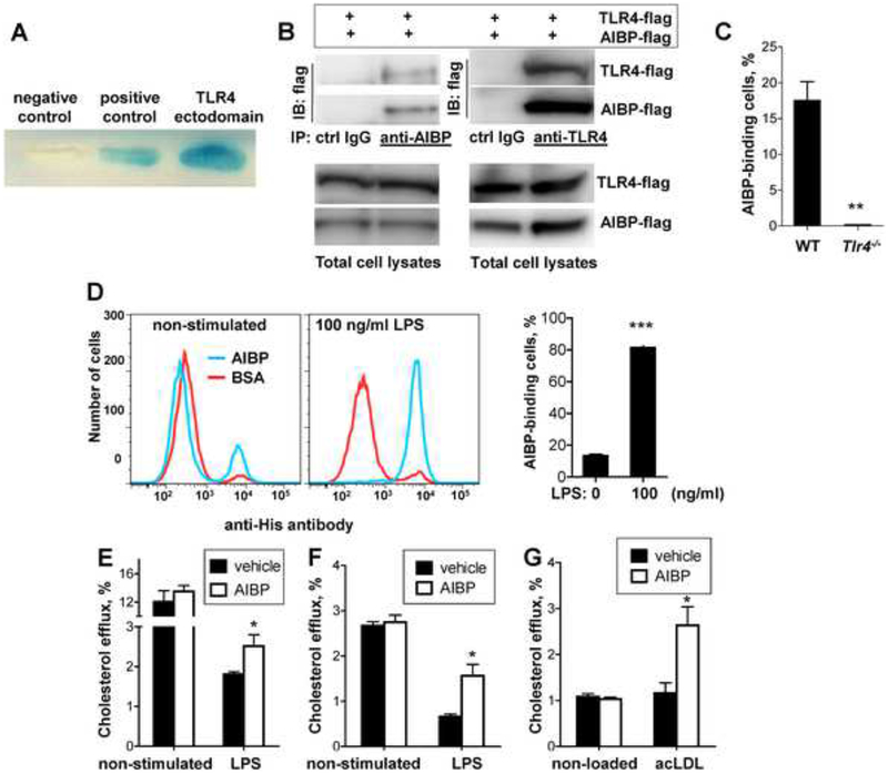
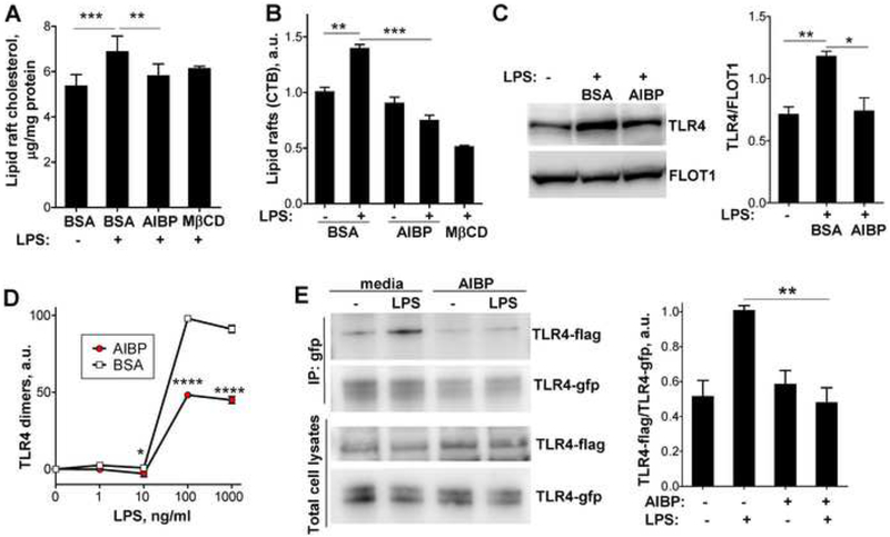
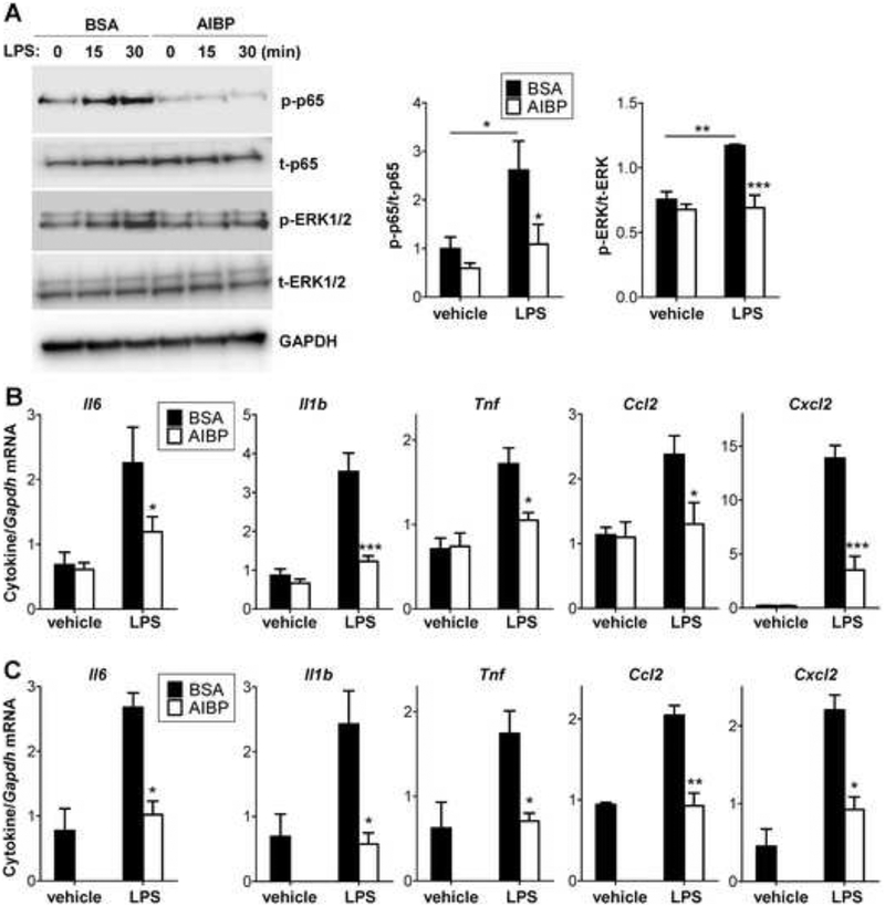
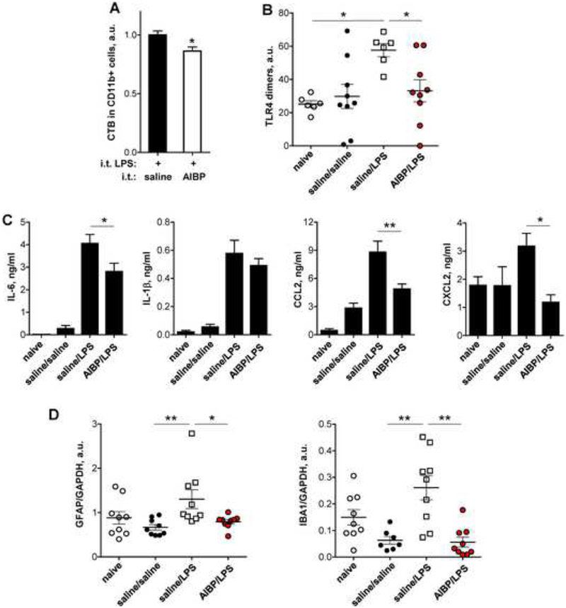
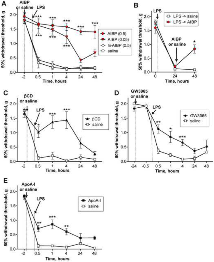
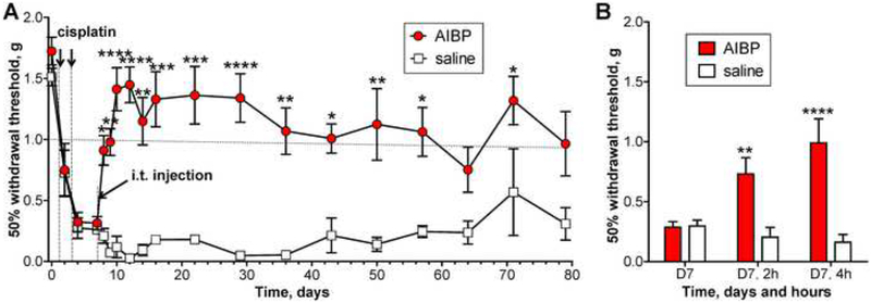
Apolipoprotein A-I binding protein (AIBP) reduces lipid raft abundance by augmenting the removal of excess cholesterol from the plasma membrane. Here, we report that AIBP prevents and reverses processes associated with neuroinflammatory-mediated spinal nociceptive processing. The mechanism involves AIBP binding to Toll-like receptor-4 (TLR4) and increased binding of AIBP to activated microglia, which mediates selective regulation of lipid rafts in inflammatory cells. AIBP-mediated lipid raft reductions downregulate LPS-induced TLR4 dimerization, inflammatory signaling, and expression of cytokines in microglia. In mice, intrathecal injections of AIBP reduce spinal myeloid cell lipid rafts, TLR4 dimerization, neuroinflammation, and glial activation. Intrathecal AIBP reverses established allodynia in mice in which pain states were induced by the chemotherapeutic cisplatin, intraplantar formalin, or intrathecal LPS, all of which are pro-nociceptive interventions known to be regulated by TLR4 signaling. These findings demonstrate a mechanism by which AIBP regulates neuroinflammation and suggest the therapeutic potential of AIBP in treating preexisting pain states.

Keywords: AIBP; TLR4; allodynia; chemotherapy; cholesterol; chronic pain; inflammation; lipid rafts; neuroinflammation; neuropathic pain.


Conflict of interest statement

DECLARATION OF INTERESTS

The authors declare no competing financial interests. Y.I.M. and T.L.Y. are inventors listed in patent applications related to the topic of this paper.

Figures

Figure 1.
Figure 1.. AIBP interaction with TLR4 and selective cholesterol efflux.
A, Yeast two-hybrid was performed with pB42AD-AIBP and pLexA-TLR4 ectodomain. The positive control was the yeast cell line EGY48/p80p-LacZ co-transfected with pLexA53 and pB42ADT; the negative control was the yeast cell line co-transfected with pLexA and pB42AD. B, HEK293 cells were co-transfected with the flag-tagged TLR4 ectodomain and flag-tagged AIBP. AIBP from cell lysates were pulled down with either anti-AIBP antibody, anti-TLR4 antibody or respective isotype control IgG. Blots of the pull-down or total cell lysates were probed with an anti-flag antibody. C, Peritoneal elicited macrophages from WT and Tlr4−/− mice were incubated for 2 hours on ice with 2 μg/ml BSA or 2 μg/ml AIBP (with a His-tag) and then subjected to a flow cytometry analysis with a FITC-conjugated anti-His antibody. D, BV-2 cells were stimulated with 100 ng/ml LPS for 15 min, placed on ice, and 2 μg/ml AIBP (His-tagged) or BSA were added for 2 hours, and cells were subjected to a flow cytometry analysis with a FITC-conjugated anti-His antibody. Mean±SEM; n=4; ***, p<0.001 (Student’s t-test). E, Primary brain microglia cells were loaded with 3H-cholesterol, equilibrated and then sequentially incubated with 0.2 μg/ml AIBP or BSA for 1 hour and 100 ng/ml LPS for 1 hour in complete medium. Cholesterol efflux was measured as described in Methods. Mean±SEM; n=3–5; *, p<0.05 (Student’s t-test). F, Human THP-1-derived macrophages were loaded with 3H-cholesterol, equilibrated and incubated for 24 hours with 3 μg/ml ApoA-I and 0.1% BSA, in the presence or absence of 0.2 μg/ml AIBP. LPS (10 μg/ml) was added during equilibration and efflux incubations. Mean±SEM; n=4; *, p<0.05 (Student’s t-test). G, Human THP-1-derived macrophages were loaded with acetylated LDL (acLDL; 50μg/ml) and 3H-cholesterol, equilibrated and incubated for 24 hours with 3 μg/ml ApoA-I and 0.1% BSA, in the presence or absence of 0.2 μg/ml AIBP. Cholesterol efflux was measured as described in Methods. Mean±SEM; n=4; *, p<0.05 (Student’s t-test). See also Figure S1A.
Figure 2.
Figure 2.. AIBP disrupts lipid rafts and inhibits TLR4 dimerization.
A-C, BV-2 cells were incubated for 2 hours with vehicle (0.1% BSA) or 0.2 μg/ml AIBP (in 0.1% BSA) in serum-containing medium and stimulated with 10 ng/ml LPS for 10 min. A, Content of free cholesterol in isolated raft fractions was normalized to total cell protein. Mean±SEM; n=6 for first 3 columns; ***, p<0.001; **, p<0.01 (repeated measures ANOVA; raft isolation was performed for a single replicate of all samples per day); n=3 for MβCD. Mean±SEM; n=3; ***, p<0.001; **, p<0.01 (one-way ANOVA). B, Content of CTB-positive lipid rafts was measured in a flow cytometry assay. C, TLR4 occupancy in isolated lipid rafts was tested in western blot. Mean±SEM; n=3; **, p<0.01; *, p<0.05 (one-way ANOVA). D, BV-2 cells were preincubated for 2 h with 0.2 μg/ml BSA or AIBP, followed by a 15 min incubation with LPS. Arbitrary numbers of TLR4 dimers were measured in a FACS assay with MTS510 and SA15–21 TLR4 antibodies as described in Methods. Mean±SD; n=3; p<0.05; ****, p<0.0001 (two-way ANOVA with Bonferroni post-test). E, Ba/F3 cells stably expressing TLR4-gfp, TLR4-flag and MD2 were incubated with serum-free media containing 50 μg/ml HDL, in the presence or absence of 0.2 μg/ml AIBP, and then stimulated with 10 ng/ml LPS for 20 min. Cell lysates were immunoprecipitated with an anti-GFP antibody and blots were probed with anti-flag and anti-GFP antibodies. Mean±SEM; n=4–6; **, p<0.01; Student’s t-test. See also Figures S1B and S2.
Figure 3.
Figure 3.. AIBP reduces inflammatory responses in microglia.
A-B, BV-2 cells were incubated for 2 hours with 0.2 μg/ml BSA or AIBP in serum-containing medium and stimulated with 10 ng/ml LPS. p65 and ERK1/2 phosphorylation were tested after 30 min (A), and cytokine mRNA expression after 2 h of incubation (B). C, Primary mouse microglia (pooled from 5–6 mice per sample) were incubated for 2 hours with 0.2 μg/ml BSA or AIBP in serum-containing medium and stimulated with 10 ng/ml LPS for 1 hour. Mean±SEM; n=4–6 for BV-2; n=3 for primary microglia experiments; *, p<0.05; **, p<0.01; ****, p<0.0005 (Student’s t-test). Due to limited availability of primary cells, ‘vehicle/AIBP’ group was omitted. See also Figures S3 and S4A.
Figure 4.
Figure 4.. Intrathecal AIBP reduces lipid rafts and TLR4 dimerization in spinal myeloid cells, neuroinflammation, and glial activation.
A-B, Male mice were given an i.t. injection of AIBP (0.5 μg/ 5 μl) or saline (5 μl); two hours later, all mice were given an i.t. injection of LPS (0.1 μg/ 5 μl) and were terminated 15 min later. A, Spinal cords were isolated, demyelinated, stained for CD11b and cholera toxin B (CTB) and subjected to a flow cytometry analysis. Mean±SEM; n=11; *, p<0.05 (Student’s t-test). B, Demyelinated spinal homogenates were stained with MTS510, SA15–21 and isotype control antibodies, analyzed by flow cytometry, and levels of TLR4 dimers were calculated as described in Methods. Mean±SEM; n=6–9; *, p<0.05 (Newman-Keuls multiple comparison test). C-D, Male mice were given an i.t. injection of AIBP (0.5 μg/ 5 μl) or saline (5 μl); two hours later, all mice were given an i.t. injection of LPS (0.1 μg/ 5 μl) and were terminated 4 hours later. Naïve mice were used as a negative control. C, CSF was isolated and tested in ELISA for the levels of inflammatory cytokines. Mean±SEM; n=8–11; *, p<0.05; **, p<0.01 (one-way ANOVA with Bonferroni’s multiple comparison test). Levels of TNFα in these samples were below detection limit. D, Lumbar spinal cord was isolated and analyzed in western blot for expression of GFAP and IBA1 (see Fig. S5). Mean±SEM; n=7–9; *, p<0.05; **, p<0.01 (non-parametric Kruskal-Wallis test with Dunn’s multiple comparison test). See also Figures S4B and S5.
Figure 5.
Figure 5.. Intrathecal AIBP prevents and reverses LPS-induced allodynia.
A, Following baseline von Frey threshold testing, male mice were given an i.t. injection of AIBP (0.05 μg/ 5 μl, n=4; or 0.5 μg/ 5 μl, n=6), heat-inactivated AIBP (hi-AIBP; 0.5 μg/ 5 μl; n=6), or saline (5 μl; n=6). B, Following baseline von Frey threshold testing, male mice were given i.t. LPS (0.1 μg/ 5 μl). Twenty-four hours later, mice received i.t. AIBP (0.5 μg/ 5 μl; n=4) or saline (5 μl; n=4). C-E, Following baseline von Frey threshold testing, male mice were given an i.t. injection of: C, beta-cyclodextrin (βCD; 5 μl of 10% solution in saline; n=4) or saline (5 μl; n=4; same group as used in panel A); D, the LXR agonist GW3965 (0.1 μg/ 5 μl; n=10) or saline (5 μl; n=6); or E, ApoA-I (5 μg/ 5 μl; n=10) or saline (5 μl; n=6). Two hours (C and E) or 24 hours (D) later, all mice were given an i.t. injection of LPS (0.1 μg/ 5 μl) and tested over time for tactile allodynia. Mean±SEM; *, p<0.05; **, p<0.01; ***, p<0.001; ****, p<0.0001 (A, C-E: two way ANOVA with Bonferroni post-test; B: Student’s t-test for the 48 hour time point only). See also Figure S6.
Figure 6.
Figure 6.. Intrathecal AIBP prevents and reverses i.t. LPS- and intraplantar formalin-induced allodynia.
A-B, Following baseline von Frey threshold testing, male mice were given an intraplantar injection of formalin in one hind paw. A, The graph shows total numbers of hind paw flinches in phase I (1–9 min) and phase II (10–50 min). Mean±SD; n=4–12 per group; #, non-significant, p>0.05 (Student’s t-test). B, The graph shows $baseline-normalized changes in the withdrawal threshold in the ipsilateral paw. Mean±SEM; n=4–12 per group; *, p<0.05 (Student’s t-test for the 7 day time point only). C, In a group of animals different from those used in panels A and B, von Frey readings were #normalized at the 7th day post-formalin and the mice received i.t. AIBP (0.5 μg/ 5 μl) or saline (5 μl). Mean±SEM; n=4 per group; *, p<0.05 between 0 and 24 hours (repeated measures one-way ANOVA with Bonferroni post-test).
Figure 7.
Figure 7.. Intrathecal AIBP reverses established cisplatin-induced allodynia.
A, Male mice received 2 i.p. injections of cisplatin (2.3 mg/kg) over a period of 3 days to establish allodynia. On day 7, mice were treated with i.t. AIBP (0.5 μg/ 5 μl) or saline (5 μl). Combined data from 2 independent experiments. B, The graph presents, in a different time scale, the experiment shown in panel A. Mice were tested on day 7 (after start of cisplatin treatment), before and 2 and 4 hours after i.t. AIBP and saline. Overall numbers of animals per group were on days 0–22: n = 17 (AIBP) and 12 (saline); days 29–79: n = 8 (AIBP) and 3 (saline). Mean±SEM. *, p<0.05; **, p<0.01; ***, p<0.001; ****, p<0.0001 (two way ANOVA with Bonferroni post-test).

References

    1. Agalave NM, Larsson M, Abdelmoaty S, Su J, Baharpoor A, Lundback P, Palmblad K, Andersson U, Harris H, and Svensson CI (2014). Spinal HMGB1 induces TLR4-mediated long-lasting hypersensitivity and glial activation and regulates pain-like behavior in experimental arthritis. Pain 155, 1802–1813. - PubMed
    1. Akashi S, Saitoh S. i., Wakabayashi Y, Kikuchi T, Takamura N, Nagai Y, Kusumoto Y, Fukase K, Kusumoto S, Adachi Y, et al. (2003). Lipopolysaccharide Interaction with Cell Surface Toll-like Receptor 4-MD-2: Higher Affinity than That with MD-2 or CD14. J Exp Med 198, 1035–1042. - PMC - PubMed
    1. Anttila V, Winsvold BS, Gormley P, Kurth T, Bettella F, McMahon G, Kallela M, Malik R, de Vries B, Terwindt G, et al. (2013). Genome-wide meta-analysis identifies new susceptibility loci for migraine. Nat Genet 45, 912–917. - PMC - PubMed
    1. Baranova I, Vishnyakova T, Bocharov A, Chen Z, Remaley AT, Stonik J, Eggerman TL, and Patterson AP (2002). Lipopolysaccharide down regulates both scavenger receptor B1 and ATP binding cassette transporter A1 in RAW cells. Infect Immun 70, 2995–3003. - PMC - PubMed
    1. Blasi E, Barluzzi R, Bocchini V, Mazzolla R, and Bistoni F (1990). Immortalization of murine microglial cells by a v-raf/v-myc carrying retrovirus. J Neuroimmunol 27, 229–237. - PubMed

Publication types

MeSH terms