. 2019 Jan;39(1):123-134.
doi: 10.1177/0333102418779557. Epub 2018 May 31.

Non-invasive dural stimulation in mice: A novel preclinical model of migraine

Affiliations

Non-invasive dural stimulation in mice: A novel preclinical model of migraine

Carolina Christina Burgos-Vega et al. Cephalalgia. 2019 Jan.

Abstract

Background: Migraine is characterized by a collection of neurological symptoms in the absence of injury or damage. However, several common preclinical migraine models require significant damage to the skull to stimulate the dura mater, the likely source of afferent signaling leading to head pain. The goal of this study was to determine whether dural stimulation can be performed in mice using an injection that does not cause injury or damage.

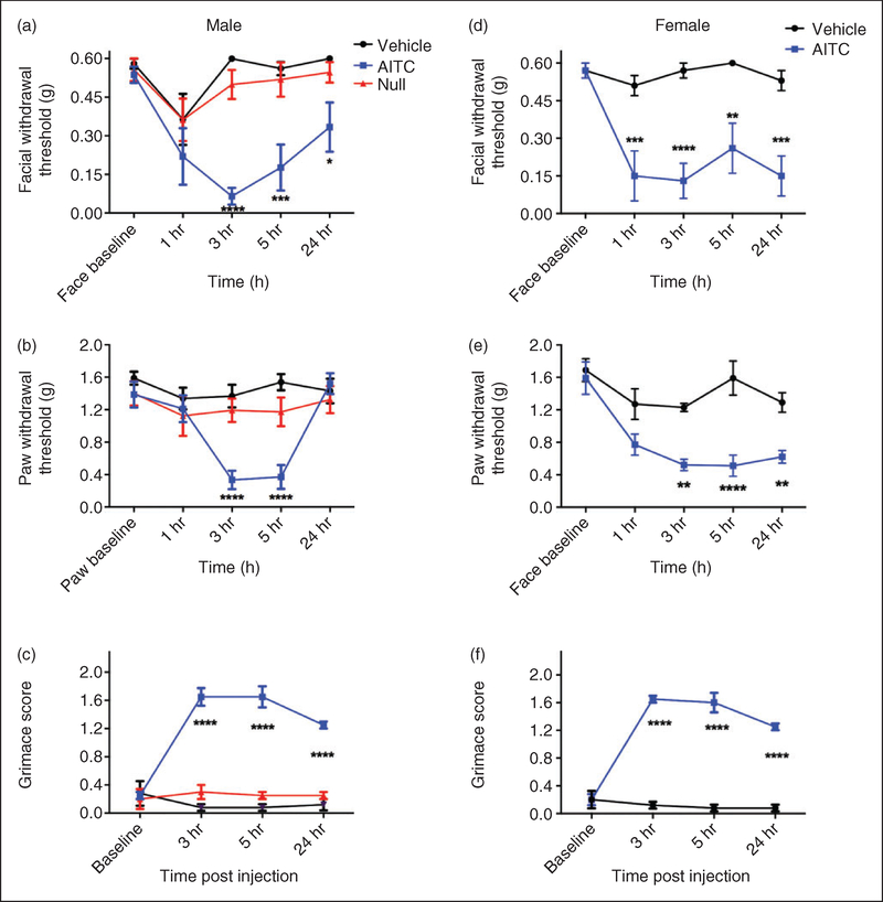
Methods: Using mice, injections of stimuli were administered to the dura mater through the soft tissue at the intersection between the lambdoidal and sagittal sutures. This technique did not require a permanent cannula nor did it cause damage to the skull or dura. Following injection of noxious stimuli, migraine-like behaviors were measured including cutaneous allodynia and facial grimace. The retrograde tracer fluorogold was applied onto the dura using the same injection technique to label trigeminal ganglion cell bodies, which were then testing in vitro using patch-clamp electrophysiology.

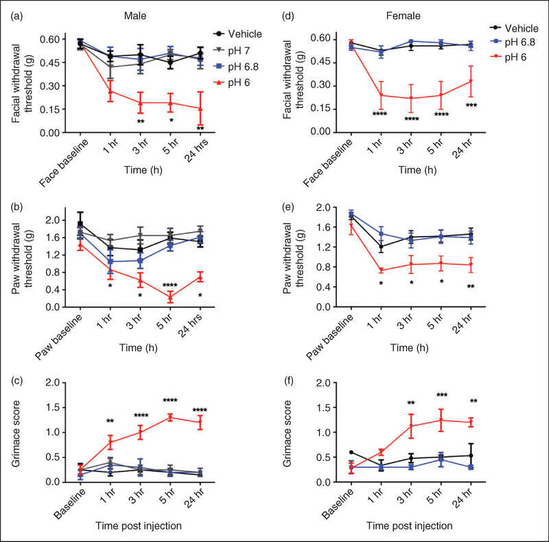
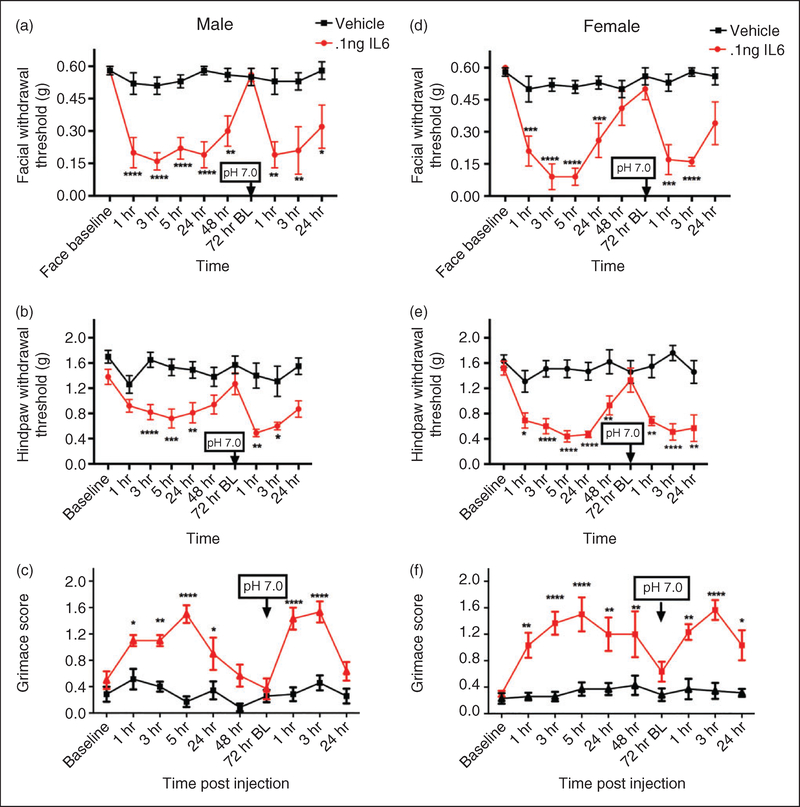
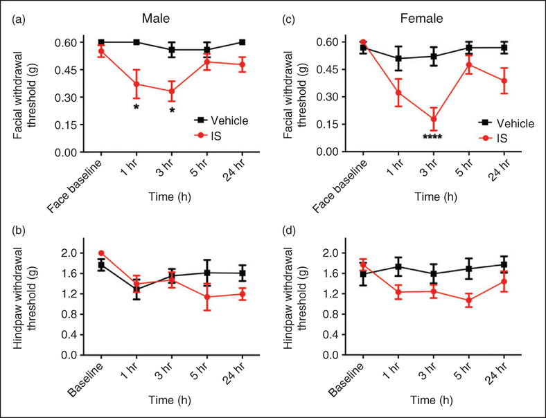
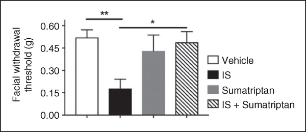
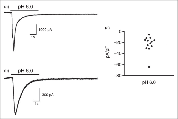
Results: Dural injection of allyl-isothiocyanate, low pH, interleukin-6, or inflammatory soup but not vehicles, led to cephalic/extracephalic allodynia. Facial grimace responses were also observed with allyl-isothiocyanate, pH 6.0, and interleukin-6. Stimulation with interleukin-6 causes priming to normally subthreshold pH 7.0 stimulation of the dura following resolution of the initial interleukin-6 behavior. Systemic injection of sumatriptan at the time of dural stimulation with inflammatory soup decreased the resulting cutaneous hypersensitivity. Trigeminal ganglion cell bodies retrogradely labeled from the dura had low pH-evoked currents similar to those generated by acid-sensing ion channels.

Conclusion: Non-invasive dural stimulation in mice can be used as a model of migraine in the absence of injury.

Keywords: Dura; headache; interleukin; meninges; migraine; mouse model; priming; trigeminal.


Conflict of interest statement

Declaration of conflicting interests

The authors declared no potential conflicts of interest with respect to the research, authorship, and/or publication of this article.

Figures

Figure 1.
Figure 1.
Non-invasive dural application of 10% AITC produces headache-related behaviors in both male and female mice. Dural application of AITC produces cutaneous allodynia in both male ((a), (b)) and female mice ((d), (e)). Withdrawal thresholds to tactile stimuli applied to the face ((a), (d)) and hindpaws ((b), (e)) were measured in animals prior to and after dural application of AITC, vehicle (mineral oil) or a null injection (n = 6 for all groups, see methods for definition of null injections). In males, null injections failed to produce significant facial and hindpaw allodynia at all timepoints ((a), (b)). Administration of AITC produced significant allodynia facial (a) and hindpaw responses (b) in males. In females, administration of AITC produced significant facial allodynia (d) and hindpaw responses (e). Grimace behaviors for both males (n = 6 all groups, (c) and females (n = 6 all groups, (f) were significant at multiple time points. Two-factor analysis of variance (ANOVA) indicated a significant effect of both treatment and time of both the face and hindpaws. Significant differences among means for each group were determined by analysis of variance followed by Bonferroni post hoc test. Males (a) facial: Time F (4, 75) = 6.602, p= 0.0001, treatment F (2, 75) = 6.602, p < 0.0001; (b) hindpaw: Time F (4, 75) = 6.572, p < 0.0001, treatment F (2, 75) = 13.03, p < 0.0001; (c) grimace: Time F (3, 40) = 11.68, P < 0.0001, treatment F (2, 40) = 131.5, P < 0.0001. Females (d) facial: Time F (4, 50) = 5.473, p= 0.001, treatment F (1, 50) = 62.11, p < 0.0001; (e) hindpaw: Time F (4, 50) = 9.213, p < 0.0001, treatment F (1, 50) = 46.4, p < 0.0001; (f) grimace: Time F (3, 28) = 26.75, p < 0.0001, treatment F (1, 28) = 303.8, p < 0.0001.
Figure 2.
Figure 2.
Non-invasive dural application of pH 6.0 produces headache-related behavior in male and female mice. Dural application of low pH (pH 6) produces cutaneous hypersensitivity in both male ((a), (b)) and female mice ((d), (e)). Withdrawal thresholds to tactile stimuli applied to the face (a) and hindpaws of male mice (b) were measured in animals prior to and following dural application of pH 6.0 (1–5 hr; n = 13, 24 hr; n = 7), pH 6.8 (n = 8) pH 7.0 (n = 7) or vehicle (pH 7.4; n = 8). In males, only administration of pH 6.0 produced significant facial (a) and hindpaw (b) responses. Similarly, female withdrawal thresholds to tactile stimuli applied to the face (d) and hindpaws (e) were measured in animals prior to and following dural application of pH 6.0 (n = 11), pH 6.8 (n = 8) or vehicle (pH 7.4, n = 16). In females, dural administration of pH 6 produced significant facial (d) and hindpaw (e) responses. Grimace behaviors were significant in males (n = 5, (c)) and in females (n = 5, (f)). Two-factor analysis of variance (ANOVA) indicated a significant effect of both treatment and time of both the face and hindpaws. Significant differences among means for each group were determined by analysis of variance followed by Bonferroni post hoc test. Males (a) facial: Time F (4, 154) = 6.631, p < 0.0001, treatment F (3, 154) = 18.12, p < 0.0001; (b) hindpaw: Time F (4, 154) = 5.7, p < 0.0001, treatment F (3, 154) = 22.75, p< 0.0001; (c) grimace: Time F (4,80) = 3.976, p= 0.00054, treatment F (3, 80) = 51.78, p < 0.0001. Females (d) facial: Time F (4, 160) = 4.64, p= 0.0014, treatment F (2, 160) = 54, p < 0.0001; (e) hindpaw: Time F (4, 160) = 11.26, p < 0.0001, treatment F (2, 160) = 22.64, p < 0.0001; (f) grimace: F (4, 60) = 4.332, p= 0.0038, treatment F (2, 60) = 24.41, p < 0.0001
Figure 3.
Figure 3.
Non-invasive dural application of IL-6 produces headache-related behavior in both male and female mice. Dural application of IL-6 (0.1 ng) produces cutaneous hypersensitivity in both male ((a), (b)) and female mice ((d), (e)). Males treated with IL-6 (1–48 hr; n = 13, 72 hr BL and later; n = 7), showed significant facial (a) and hindpaw (b) responses and also significant responses to subsequent dural pH 7.0. Similarly, females treated with IL-6/dural pH 7.0 (1–48 hr; n = 12, 72 hr BL and later; n = 7) had significant facial (d), and hindpaw (e) responses and also significant responses to subsequent dural pH 7.0. Grimace behaviors were significant in males (n = 6, all timepoints; (c) and in females (n = 7, (e)). Significant differences among means for each group were determined with one-way ANOVA followed by Bonferroni post hoc test. Males facial: Time F (9, 177) = 4.768, p < 0.0001, treatment F (1, 177) = 106.5, p < 0.0001; hindpaw: Time F (9, 166) = 3.135, p= 0.0016, treatment F (1, 166) = 82.65, p < 0.0001); grimace: Time F (9, 110) = 7.305, p < 0.0001, treatment F (1, 110) = 127.4, p < 0.0001. Females facial: Time F (9, 151) = 6.818, p < 0.0001, treatment F (1, 151) = 120.7, p < 0.0001; hindpaw: Time F (9, 151) = 3.758, p < 0.0001, treatment F (1, 151) = 130.4, p < 0.0001); grimace: Time F (9, 110) = 3.578, p= 0.0004, treatment F (1, 110) = 125.8, p < 0.0001.
Figure 4.
Figure 4.
Non-invasive dural application of IS produces headache-related behavior in both male and female mice. Dural application of IS produces cutaneous facial hypersensitivity in both male (a) and female mice (d). Mice treated with IS (1–24 hr; n = 6–10), showed significant facial (a) and (c) responses, but no significant differences in hindpaw sensitivity were observed ((b) and (d)). Significant differences among means for each group were determined by two-way ANOVA followed by Bonferroni post hoc test. Males facial: Time F (4, 70) = 1.95, p= 0.1117, treatment F (1, 70) = 18.54, p < 0.0001; hindpaw: Time F (4, 55) = 2.903, p= 0.0299, treatment F (1, 55) = 1.128, p= 0.2928. Females facial: Time F (4, 75) = 5.908, p= 0.0003, treatment F (1, 75) = 21.28, p < 0.0001; hindpaw: Time F (4, 60) = 1.111, p= 0.3599, treatment F (1, 60) = 9.261, p= 0.0035.
Figure 5.
Figure 5.
Cutaneous facial allodynia following non-invasive dural application of IS is blocked by sumatriptan. Dural application of IS produced cutaneous facial hypersensitivity in female mice. Withdrawal thresholds to tactile stimuli applied to the face were measured in mice at the 3-hour time point following dural application of vehicle, IS, sumatriptan, or IS + sumatriptan (n = 6–9). A significant decrease in withdrawal threshold was observed only in the IS group. Significant differences among means for each group were determined by analysis of variance followed by Bonferroni post hoc test. Females facial: Treatment F (3, 26) = 5.408, p= 0.005.
Figure 6.
Figure 6.
Mouse trigeminal ganglion neurons retrogradely labeled from the dura mater generate ASIC-like currents at pH 6.0. Recordings from retrogradely-labeled mouse dural afferents in response to a 5-sec pH change from 7.4 to 6.0 shown in (a) and (b) are characteristic of ASIC currents. (c) Current density (pA/pF) of the 13 neurons that responded to pH 6.0 (out of 20 total neurons recorded) range from 5–65 pA/pF with a mean of 22.5 pA/pF.

References

    1. Hougaard A, Amin FM and Ashina M. Migraine and structural abnormalities in the brain. Curr Opin Neurol 2014; 27: 309–314. - PubMed
    1. Burstein R, Noseda R and Borsook D. Migraine: Multiple processes, complex pathophysiology. J Neurosci 2015; 35: 6619–6629. - PMC - PubMed
    1. Goadsby PJ, Holland PR, Martins-Oliveira M, et al. Pathophysiology of migraine: A disorder of sensory processing. Physiol Rev 2017; 97: 553–622. - PMC - PubMed
    1. Vos T, Flaxman AD, Naghavi M, et al. Years lived with disability (YLDs) for 1160 sequelae of 289 diseases and injuries 1990–2010: A systematic analysis for the Global Burden of Disease Study 2010. Lancet 2012; 380: 2163–2196. - PMC - PubMed
    1. Diener HC, Dodick DW, Goadsby PJ, et al. Chronic migraine – classification, characteristics and treatment. Nat Rev Neurol 2012; 8: 162–171. - PubMed