Review
. 2018 May 30;18(1):32.
doi: 10.1186/s12893-018-0365-8.

Para-duodenal hernia: a report of five cases and review of literature

Affiliations
Review

Para-duodenal hernia: a report of five cases and review of literature

Kamleshsingh Shadhu et al. BMC Surg. .

Abstract

Background: Para-duodenal hernia (PDH) represents rare clinical entities based on few literatures.

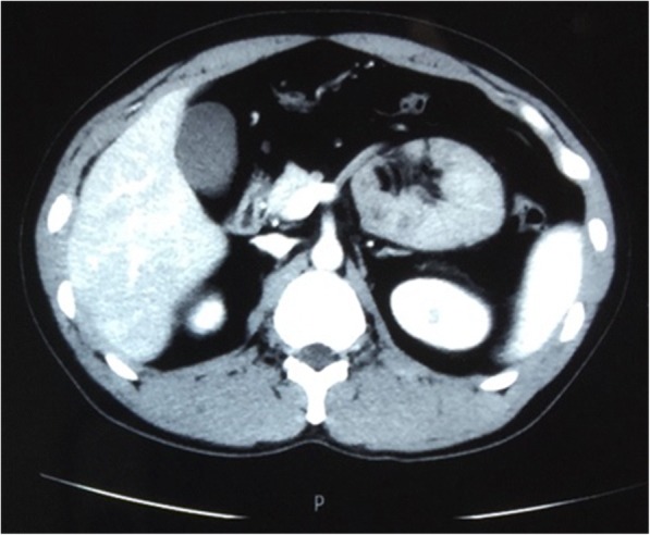
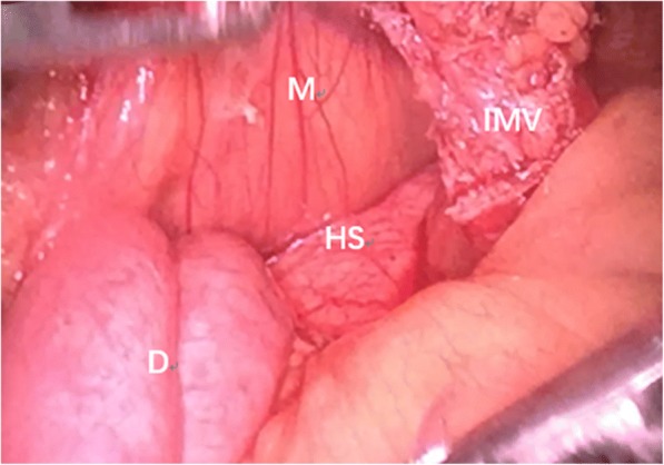
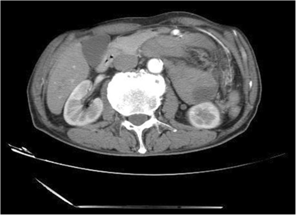
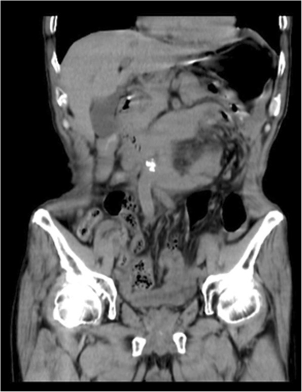
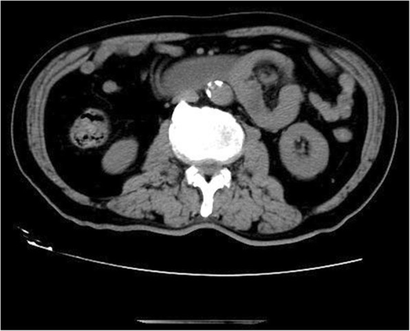
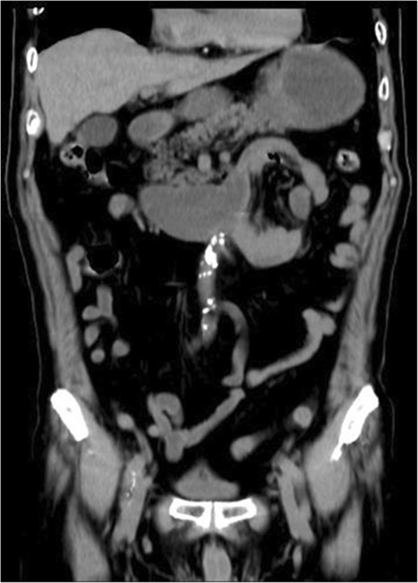
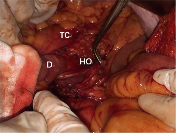
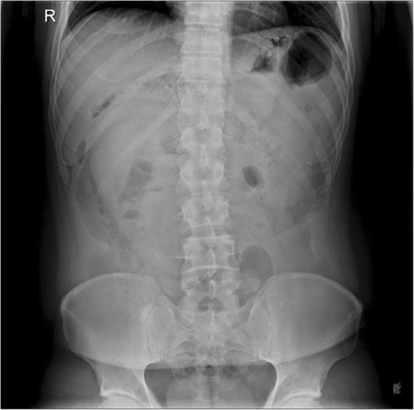
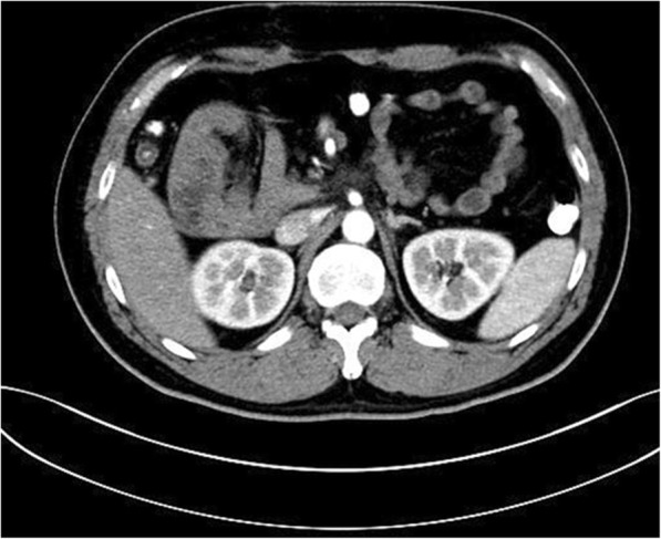
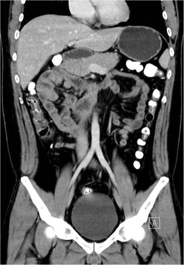
Case presentation: We report five cases of Para-duodenal hernia, all occurring in male patients ranging from 34 to 75 years of age. The patients had varied manifestations presenting with abdominal pain with or without vomiting and nausea and with or without signs of intestinal obstruction. CT images showed cluster of dilated bowel segments with displaced mesenteric vessels at hernial orifice. Laparoscopic surgical approach was adopted, and the patients were discharged about a week later without further complications.

Conclusion: We hope to raise awareness about the management of this rare clinical entity and the benefits of CT imaging and laparoscopic surgery as standard approaches.

Keywords: Abdominal pain; CT images; Intestinal obstruction; Laparoscopic surgery; Paraduodenal hernia.


Conflict of interest statement

Ethics approval and consent to participate

Not applicable.

Consent for publication

Written informed consent was obtained from the participants for publication of this article and any accompanying tables/images. A copy of the written consent is available for review by the Editor of this journal.

Competing interests

The authors declare that they have no competing interests.

Publisher’s Note

Springer Nature remains neutral with regard to jurisdictional claims in published maps and institutional affiliations.

Figures

Fig. 1
Fig. 1
Transverse view of CT image showing the cluster of small intestine and its mesentery. The entrapped intestinal loop was behind the infernal mesenteric vein (white arrow)
Fig. 2
Fig. 2
The defect in the transverse mesocolon. M: transverse mesocolon, D: the ascending part of duodenum, HO: hernia orifice
Fig. 3
Fig. 3
Transverse view of CT image showing the cluster of small intestine and its mesentery
Fig. 4
Fig. 4
The defect in the transverse mesocolon was opened. M: mesocolon, IMV: inferior mesenteric vein, D: the ascending part of duodenum, HS: hernia sac
Fig. 5
Fig. 5
Transverse view of CT image showing the cluster of small intestine and its mesentery entrapped in the left side of abdomen, beneath the left branch of middle mesenteric artery. The attenuated enhancement of the entrapped intestinal loop suggested intestinal ischemia
Fig. 6
Fig. 6
Coronary view of CT scan image showing the cluster of the small intestine
Fig. 7
Fig. 7
The defect in the transverse mesocolon. TC: transverse colon, D: the ascending part of duodenum, HO: hernia orifice
Fig. 8
Fig. 8
Transverse section of CT scan image showing the cluster of jejunum
Fig. 9
Fig. 9
Coronary view of CT scan image showing the cluster of the small intestine
Fig. 10
Fig. 10
The defect of transverse mesocolon was identified and closed with interrupted sutures. TC: transverse colon, D: the ascending part of duodenum, HO: hernia orifice
Fig. 11
Fig. 11
Abdominal X-ray showed a mass in the right upper quadrant of abdomen
Fig. 12
Fig. 12
Transverse view of CT image showing the cluster of small intestine and its mesentery on the right upper quadrant of abdomen before the descending part of duodenum
Fig. 13
Fig. 13
Coronary view of CT image showing the cluster of small intestine and its mesentery trapped within the right side of mesocolon

References

    1. Blachar A, Federle MP, Dodson SF. Internal hernia: clinical and imaging findings in 17 patients with emphasis on CT criteria. Radiology. 2001;218(1):68–74. doi: 10.1148/radiology.218.1.r01ja5368. - DOI - PubMed
    1. Khan MA, Lo AY, Vande Maele DM. Paraduodenal hernia. Am Surg. 1998;64(12):1218–1222. - PubMed
    1. Husain A, et al. Internal Hernia through Paraduodenal Recess with Acute Intestinal Obstruction: A Case Report. Indian J Surg. 2012;74(4):354–355. doi: 10.1007/s12262-011-0243-4. - DOI - PMC - PubMed
    1. Zonca P, et al. Treitz’s hernia. Hernia. 2008;12(5):531–534. doi: 10.1007/s10029-008-0339-3. - DOI - PubMed
    1. Armstrong O, et al. Internal hernias: anatomical basis and clinical relevance. Surg Radiol Anat. 2007;29(4):333–337. doi: 10.1007/s00276-007-0212-6. - DOI - PubMed