

Lateral line placodes of aquatic vertebrates are evolutionarily conserved in mammals
- PMID: 29848488
- PMCID: PMC6031350
- DOI: 10.1242/bio.031815
Lateral line placodes of aquatic vertebrates are evolutionarily conserved in mammals
Abstract
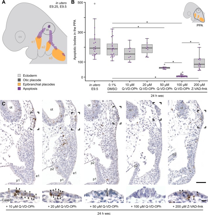
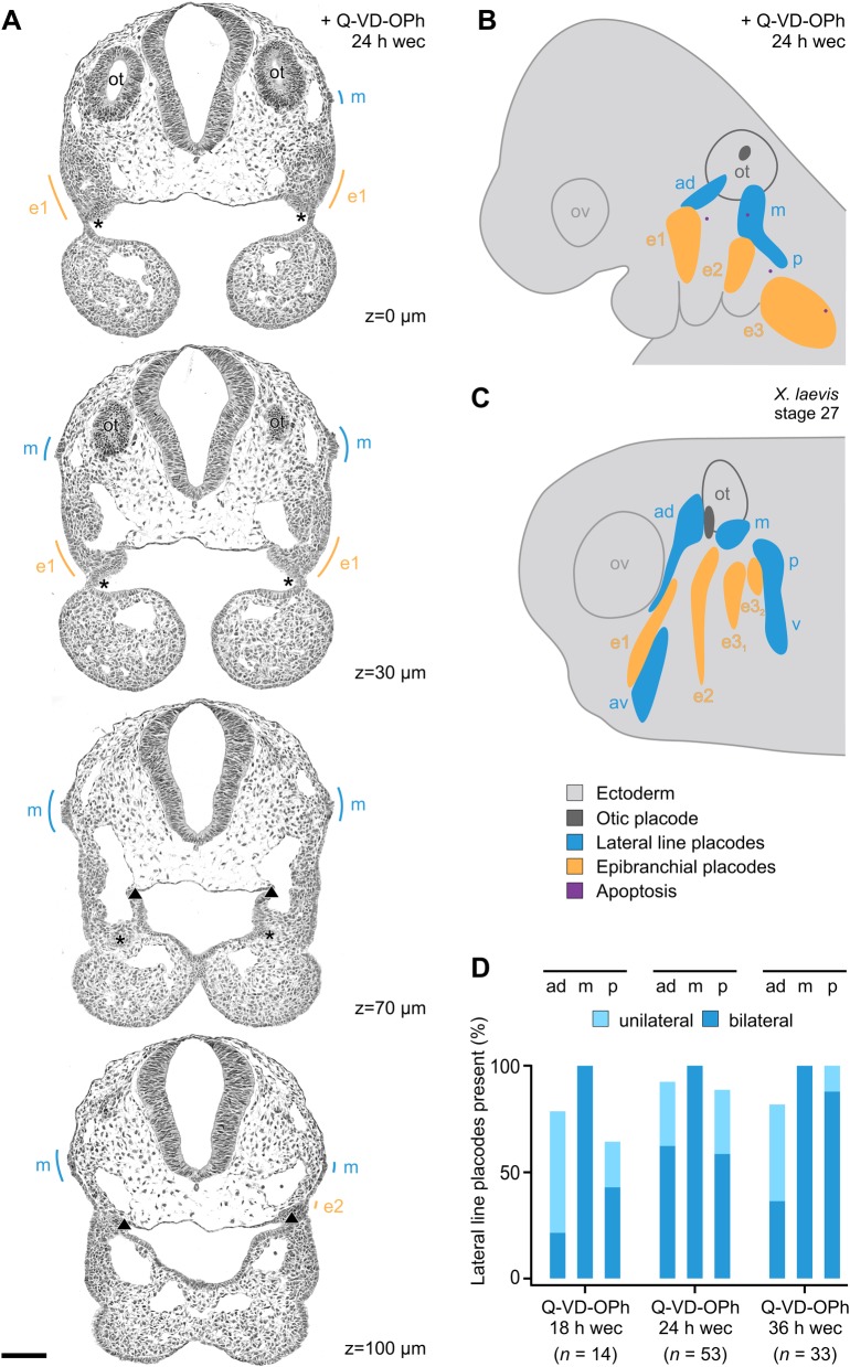
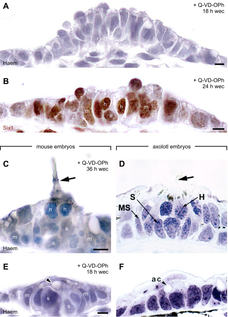
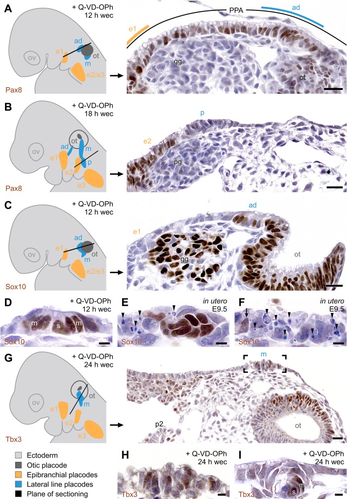
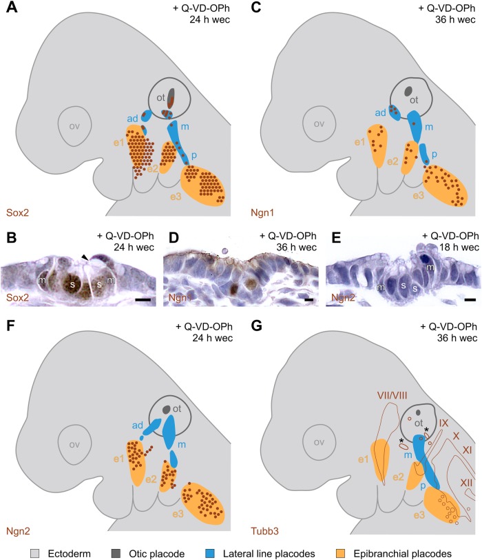
Placodes are focal thickenings of the surface ectoderm which, together with neural crest, generate the peripheral nervous system of the vertebrate head. Here we examine how, in embryonic mice, apoptosis contributes to the remodelling of the primordial posterior placodal area (PPA) into physically separated otic and epibranchial placodes. Using pharmacological inhibition of apoptosis-associated caspases, we find evidence that apoptosis eliminates hitherto undiscovered rudiments of the lateral line sensory system which, in fish and aquatic amphibia, serves to detect movements, pressure changes or electric fields in the surrounding water. Our results refute the evolutionary theory, valid for more than a century that the whole lateral line was completely lost in amniotes. Instead, those parts of the PPA which, under experimental conditions, escape apoptosis have retained the developmental potential to produce lateral line placodes and the primordia of neuromasts that represent the major functional units of the mechanosensory lateral line system.
Keywords: Apoptosis; Lateral line; Mouse embryos; Neuromasts; Posterior placodal area; Vestigial placodes.
© 2018. Published by The Company of Biologists Ltd.
Conflict of interest statement
Competing interestsThe authors declare no competing or financial interests.
Figures
Similar articles
-
Chicken embryos share mammalian patterns of apoptosis in the posterior placodal area.J Anat. 2019 Apr;234(4):551-563. doi: 10.1111/joa.12945. Epub 2019 Feb 7. J Anat. 2019. PMID: 30734277 Free PMC article.
-
The evolutionary history of vertebrate cranial placodes II. Evolution of ectodermal patterning.Dev Biol. 2014 May 1;389(1):98-119. doi: 10.1016/j.ydbio.2014.01.019. Epub 2014 Feb 1. Dev Biol. 2014. PMID: 24491817 Review.
-
Pax2/Pax8-defined subdomains and the occurrence of apoptosis in the posterior placodal area of mice.Brain Struct Funct. 2017 Aug;222(6):2671-2695. doi: 10.1007/s00429-016-1364-0. Epub 2017 Feb 3. Brain Struct Funct. 2017. PMID: 28160066
-
Induction of the epibranchial placodes.Development. 1999 Feb;126(5):895-902. doi: 10.1242/dev.126.5.895. Development. 1999. PMID: 9927591
-
Lateral line, otic and epibranchial placodes: developmental and evolutionary links?J Exp Zool B Mol Dev Evol. 2008 Jun 15;310(4):370-83. doi: 10.1002/jez.b.21188. J Exp Zool B Mol Dev Evol. 2008. PMID: 17638322 Free PMC article. Review.
Cited by
-
Evolutionary and Developmental Associations of Neural Crest and Placodes in the Vertebrate Head: Insights From Jawless Vertebrates.Front Physiol. 2020 Aug 13;11:986. doi: 10.3389/fphys.2020.00986. eCollection 2020. Front Physiol. 2020. PMID: 32903576 Free PMC article. Review.
-
Chicken embryos share mammalian patterns of apoptosis in the posterior placodal area.J Anat. 2019 Apr;234(4):551-563. doi: 10.1111/joa.12945. Epub 2019 Feb 7. J Anat. 2019. PMID: 30734277 Free PMC article.
-
Notch signalling regulates epibranchial placode patterning and segregation.Development. 2020 Feb 17;147(4):dev183665. doi: 10.1242/dev.183665. Development. 2020. PMID: 31988190 Free PMC article.
-
Senescence and Apoptosis: Architects of Mammalian Development.Front Cell Dev Biol. 2021 Jan 18;8:620089. doi: 10.3389/fcell.2020.620089. eCollection 2020. Front Cell Dev Biol. 2021. PMID: 33537310 Free PMC article. Review.
-
Programmed Cell Death Not as Sledgehammer but as Chisel: Apoptosis in Normal and Abnormal Craniofacial Patterning and Development.Front Cell Dev Biol. 2021 Oct 8;9:717404. doi: 10.3389/fcell.2021.717404. eCollection 2021. Front Cell Dev Biol. 2021. PMID: 34692678 Free PMC article. Review.
References
-
- Adameyko I., Lallemend F., Furlan A., Zinin N., Aranda S., Kitambi S. S., Blanchart A., Favaro R., Nicolis S., Lübke M. et al. (2012). Sox2 and Mitf cross-regulatory interactions consolidate progenitor and melanocyte lineages in the cranial neural crest. Development 139, 397-410. 10.1242/dev.065581 - DOI - PMC - PubMed
-
- Akazawa C., Ishibashi M., Shimizu C., Nakanishi S. and Kageyama R. (1995). A mammalian helix-loop-helix factor structurally related to the product of Drosophila proneural gene atonal is a positive transcriptional regulator expressed in the developing nervous system. J. Biol. Chem. 270, 8730-8738. 10.1074/jbc.270.15.8730 - DOI - PubMed
LinkOut - more resources
Full Text Sources
Other Literature Sources
