. 2018 Sep 15;24(18):4416-4428.
doi: 10.1158/1078-0432.CCR-17-3649. Epub 2018 May 30.

Prospective Analysis of Adoptive TIL Therapy in Patients with Metastatic Melanoma: Response, Impact of Anti-CTLA4, and Biomarkers to Predict Clinical Outcome

Affiliations

Prospective Analysis of Adoptive TIL Therapy in Patients with Metastatic Melanoma: Response, Impact of Anti-CTLA4, and Biomarkers to Predict Clinical Outcome

Marie-Andrée Forget et al. Clin Cancer Res. .

Abstract

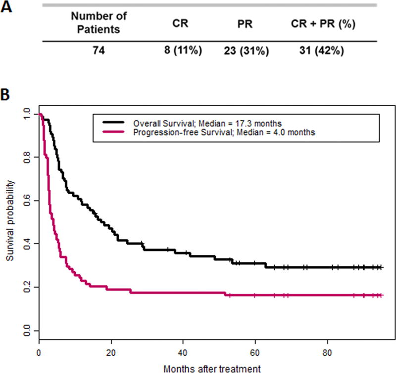
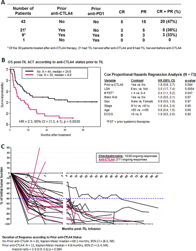
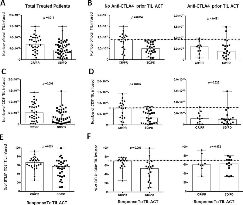
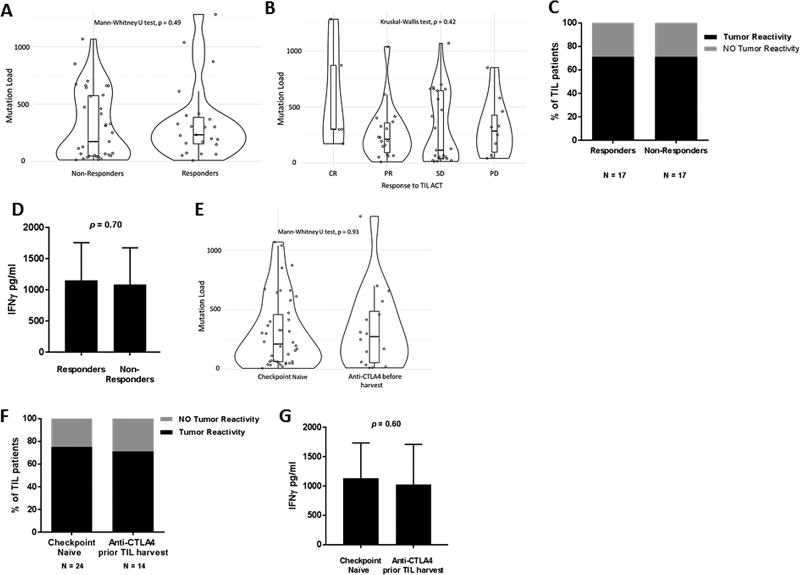
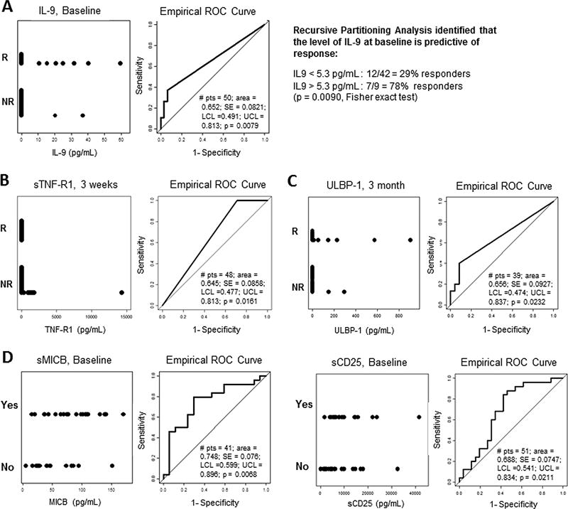
Purpose: Adoptive cell therapy (ACT) using tumor-infiltrating lymphocytes (TIL) has consistently demonstrated clinical efficacy in metastatic melanoma. Recent widespread use of checkpoint blockade has shifted the treatment landscape, raising questions regarding impact of these therapies on response to TIL and appropriate immunotherapy sequence.Patients and Methods: Seventy-four metastatic melanoma patients were treated with autologous TIL and evaluated for clinical response according to irRC, overall survival, and progression-free survival. Immunologic factors associated with response were also evaluated.Results: Best overall response for the entire cohort was 42%; 47% in 43 checkpoint-naïve patients, 38% when patients were exposed to anti-CTLA4 alone (21 patients) and 33% if also exposed to anti-PD1 (9 patients) prior to TIL ACT. Median overall survival was 17.3 months; 24.6 months in CTLA4-naïve patients and 8.6 months in patients with prior CTLA4 blockade. The latter patients were infused with fewer TIL and experienced a shorter duration of response. Infusion of higher numbers of TIL with CD8 predominance and expression of BTLA correlated with improved response in anti-CTLA4 naïve patients, but not in anti-CTLA4 refractory patients. Baseline serum levels of IL9 predicted response to TIL ACT, while TIL persistence, tumor recognition, and mutation burden did not correlate with outcome.Conclusions: This study demonstrates the deleterious effects of prior exposure to anti-CTLA4 on TIL ACT response and shows that baseline IL9 levels can potentially serve as a predictive tool to select the appropriate sequence of immunotherapies. Clin Cancer Res; 24(18); 4416-28. ©2018 AACR.


Conflict of interest statement

Conflict of interest disclosure: The authors have no competing interest to disclose.

Figures

Figure 1
Figure 1. Best overall response, OS and PFS of 74 metastatic melanoma patients following TIL ACT
(A) Clinical response is stratified by complete response (CR) and partial response (PR). (B) Overall survival (black line) and progression-free survival (pink line) post-TIL ACT of the same cohort.
Figure 2
Figure 2. Impact of checkpoint blockade exposure prior to TIL ACT on response and survival
(A) Best overall response stratified by checkpoint blockade therapy (anti-CTLA4 and/or anti-PD1) prior to TIL ACT (n=74). (B) On the left side, overall survival of checkpoint naïve patients prior to TIL ACT (black line) and anti-CTLA4 exposed patients prior to TIL (pink line). On the right side, multivariable analysis including 8 parameters. (C) Spider plot illustrating tumor regression through time after TIL infusion for checkpoint naïve patients prior to TIL ACT (black line) and anti-CTLA4 exposed patients prior to TIL (pink line). Only responders to TIL ACT are represented.
Figure 3
Figure 3. Exposure to anti-CTLA does not impact TIL persistence
(A) Assessment of TIL diversity in the infusion product at the clonal level from tumor harvested pre- (yellow) and post-(blue) anti-CTLA4 therapy for 2 patients. The clonal diversity is expressed by the clonal index (1 = a clonal population, a clonal index closer to 0 = a polyclonal population). (B) Venn diagrams depicting the number of clones found only in one of the 2 conditions [pre (yellow) or post (blue)] as well as the number of REP expanded clones found in both pre and post-anti-CTLA4 TIL products combined in the infusion product (2 patients). (C) Separate tracking TIL post-infusion of both pre- (yellow) and post- (blue) anti-CTLA4 therapy product in both patients over time. For each patients, the percentage change in the tumor burden, baseline being “0” and a negative percentage demonstrating a response.
Figure 4
Figure 4. Infusion product parameters correlating with clinical response
(A) Total amount of TIL infused per patient (n=74) stratified by responders (CR, PR) and non-responders (SD, PD). (B) Results reported in A) separated according to their status prior to TIL ACT: exposed to anti-CTLA4 (right) or checkpoint naïve (left). (C) Total amount of CD8+ TIL infused per patient stratified by clinical response. (D) Results reported in (C) separated according to their status prior to TIL ACT: exposed to anti-CTLA4 (right) or checkpoint naïve (left). (E) Percentage of CD8+BTLA+ TIL infused per patient stratified by clinical response. This percentage was evaluated by flow cytometry. (F) Results reported in (E) separated according to their status prior to TIL ACT: exposed to anti-CTLA4 (right) or checkpoint naïve (left). All P-values were generated with the Mann-Whitney test.
Figure 5
Figure 5. Mutation load and tumor recognition are not affected by exposure to anti-CTLA4
(A–B) Non-synonymous mutation load obtained from whole genome sequencing (WES) from tumor utilized for TIL propagation of treated patient. The graphs compare the mutation load from responders and non-responders (A) and from stratifying these 2 categories: CR and PR for responders and SD and PD for non-responders (B). (C) Pre-REP TIL from treated patients were co-cultured with autologous tumor targets and assessed for IFNg production by ELISA. The percentage of patients displaying tumor recognition (>200 pg/mL) is shown in black. (D) The average of IFNg secreted by the TIL lines in both group of patients capable of autologous tumor recognition shown in (C). (E) Non-synonymous mutation load comparing checkpoint naive tumors vs anti-CTLA4 exposed tumors. (F) Pre-REP TIL (regardless of patient receiving TIL therapy) were co-cultured and assessed for IFNg production as mentioned in (C). (G) The average of IFNg secreted by the TIL lines in both group of patients capable of autologous tumor shown in (F).
Figure 6
Figure 6. Correlation of serum biomarkers with response to TIL ACT and anti-CTLA4 exposure
(A–D) Grouped dot plot (left graph) and ROC curve (right graph) of the different time points in the serum of TIL treated patients. (A) Baseline serum level of circulating IL-9 comparing responders (R) from non-responders (NR) (n=50). Using recursive partitioning analysis, baseline IL-9 levels are predictive of response to TIL ACT. (B) Pre-IL-2 serum level of sTNF-R1 comparing responders (R) from non-responders (NR) (second round of IL-2, ~ 3 weeks post-TIL infusion) (n=48). (C) 3 month post-infusion level of sULBP-1 comparing responders (R) from non-responders (NR) (second round of IL-2, ~3 weeks post-TIL infusion) (n=39). (D) Baseline serum level of sMICB (n=41) and sCD25 (n=51) of patients exposed to anti-CTLA4 (YES) or not (NO) prior to TIL ACT. Number of patient in each groups varies depending on availably for either time point or marker panel.

References

    1. Dudley ME, Wunderlich JR, Robbins PF, Yang JC, Hwu P, Schwartzentruber DJ, et al. Cancer regression and autoimmunity in patients after clonal repopulation with antitumor lymphocytes. Science. 2002;298(5594):850–4. doi: 10.1126/science.1076514. - DOI - PMC - PubMed
    1. Radvanyi LG, Bernatchez C, Zhang M, Fox PS, Miller P, Chacon J, et al. Specific lymphocyte subsets predict response to adoptive cell therapy using expanded autologous tumor-infiltrating lymphocytes in metastatic melanoma patients. Clin Cancer Res. 2012;18(24):6758–70. doi: 10.1158/1078-0432.CCR-12-1177. - DOI - PMC - PubMed
    1. Besser MJ, Shapira-Frommer R, Itzhaki O, Treves AJ, Zippel DB, Levy D, et al. Adoptive transfer of tumor-infiltrating lymphocytes in patients with metastatic melanoma: intent-to-treat analysis and efficacy after failure to prior immunotherapies. Clin Cancer Res. 2013;19(17):4792–800. doi: 10.1158/1078-0432.CCR-13-0380. - DOI - PubMed
    1. Andersen R, Donia M, Ellebaek E, Borch TH, Kongsted P, Iversen TZ, et al. Long-lasting complete responses in patients with metastatic melanoma after adoptive cell therapy with tumor-infiltrating lymphocytes and an attenuated IL-2 regimen. Clin Cancer Res. 2016 doi: 10.1158/1078-0432.CCR-15-1879. - DOI - PubMed
    1. Goff SL, Dudley ME, Citrin DE, Somerville RP, Wunderlich JR, Danforth DN, et al. Randomized, Prospective Evaluation Comparing Intensity of Lymphodepletion Before Adoptive Transfer of Tumor-Infiltrating Lymphocytes for Patients With Metastatic Melanoma. J Clin Oncol. 2016;34(20):2389–97. doi: 10.1200/JCO.2016.66.7220. - DOI - PMC - PubMed

Publication types

MeSH terms