. 2018 Jul 31;92(16):e00769-18.
doi: 10.1128/JVI.00769-18. Print 2018 Aug 15.

Smc5/6 Antagonism by HBx Is an Evolutionarily Conserved Function of Hepatitis B Virus Infection in Mammals

Affiliations

Smc5/6 Antagonism by HBx Is an Evolutionarily Conserved Function of Hepatitis B Virus Infection in Mammals

Fabien Abdul et al. J Virol. .

Abstract

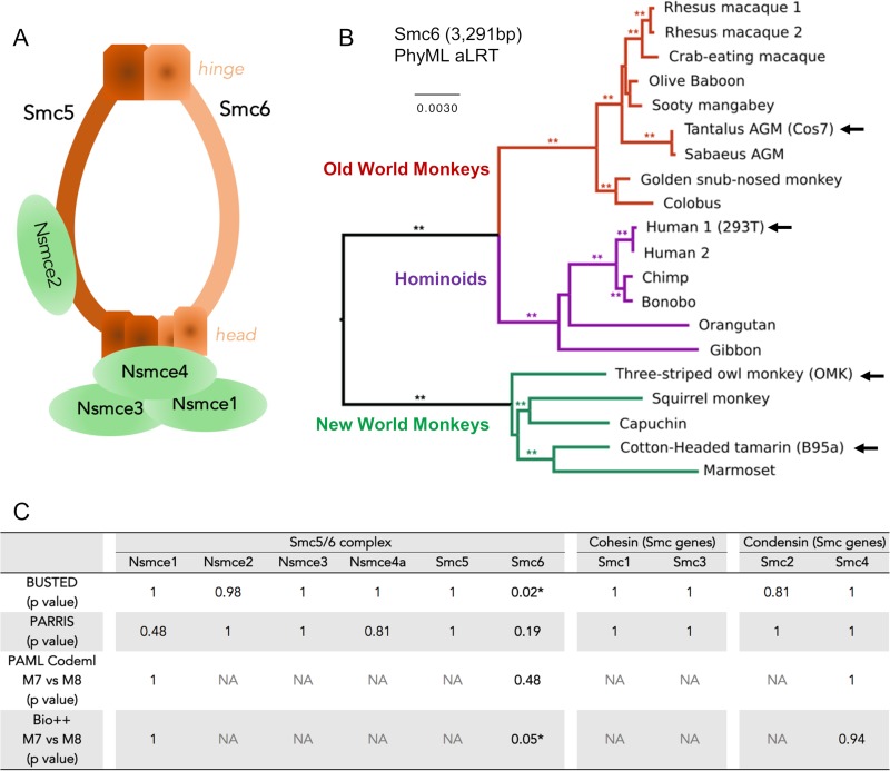
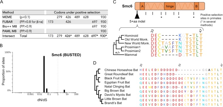
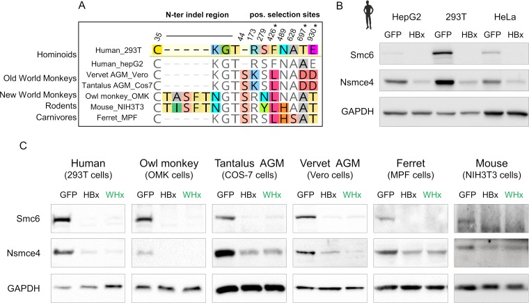
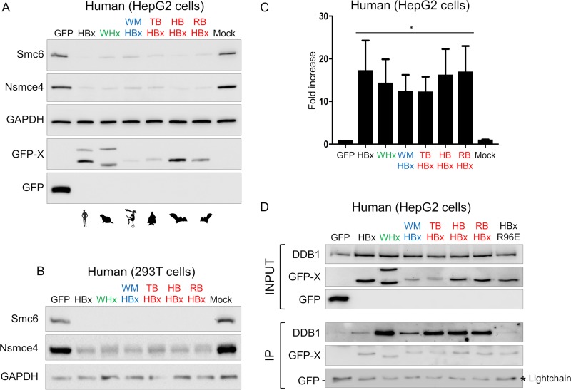
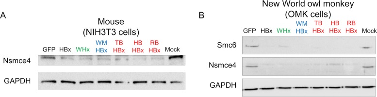
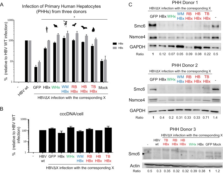
Chronic infection with hepatitis B virus (HBV) is a major cause of liver disease and cancer in humans. HBVs (family Hepadnaviridae) have been associated with mammals for millions of years. Recently, the Smc5/6 complex, known for its essential housekeeping functions in genome maintenance, was identified as an antiviral restriction factor of human HBV. The virus has, however, evolved to counteract this defense mechanism by degrading the complex via its regulatory HBx protein. Whether the antiviral activity of the Smc5/6 complex against hepadnaviruses is an important and evolutionarily conserved function is unknown. In this study, we used an evolutionary and functional approach to address this question. We first performed phylogenetic and positive selection analyses of the Smc5/6 complex subunits and found that they have been conserved in primates and mammals. Yet, Smc6 showed marks of adaptive evolution, potentially reminiscent of a virus-host "arms race." We then functionally tested the HBx proteins from six divergent hepadnaviruses naturally infecting primates, rodents, and bats. We demonstrate that despite little sequence homology, these HBx proteins efficiently degraded mammalian Smc5/6 complexes, independently of the host species and of the sites under positive selection. Importantly, all HBx proteins also rescued the replication of an HBx-deficient HBV in primary human hepatocytes. These findings point to an evolutionarily conserved requirement for Smc5/6 inactivation by HBx, showing that Smc5/6 antiviral activity has been an important defense mechanism against hepadnaviruses in mammals. It will be interesting to investigate whether Smc5/6 may further be a restriction factor of other, yet-unidentified viruses that may have driven some of its adaptation.IMPORTANCE Infection with hepatitis B virus (HBV) led to 887,000 human deaths in 2015. HBV has been coevolving with mammals for millions of years. Recently, the Smc5/6 complex, which has essential housekeeping functions, was identified as a restriction factor of human HBV antagonized by the regulatory HBx protein. Here we address whether the antiviral activity of Smc5/6 is an important evolutionarily conserved function. We found that all six subunits of Smc5/6 have been conserved in primates, with only Smc6 showing signatures of an "evolutionary arms race." Using evolution-guided functional analyses that included infections of primary human hepatocytes, we demonstrated that HBx proteins from very divergent mammalian HBVs could all efficiently antagonize Smc5/6, independently of the host species and sites under positive selection. These findings show that Smc5/6 antiviral activity against HBV is an important function in mammals. They also raise the intriguing possibility that Smc5/6 may restrict other, yet-unidentified viruses.

Keywords: HBx; Smc5/6 complex; antagonism; evolution of virus and host genes; hepatitis B virus; positive selection; restriction factor; virus-host interaction.


Figures

FIG 1
FIG 1
Smc6 is the least conserved subunit of the Smc5/6 complex in primates. (A) Architecture of the Smc5/6 complex. The complex is made of two core subunits (Smc5 and Smc6) and four non-SMC elements (Nsmce1 to Nsmce4). (B) Phylogenetic analysis of primate Smc6 genes. Sequences were aligned with MUSCLE and phylogeny was performed with PhyML and an HKY+I+G model with an approximate likelihood ratio test (aLRT) as statistical support (**, aLRT > 0.8). Newly sequenced genes (arrow) are indicated. The newly sequenced Smc6 gene from Chlorocebus pygerythrus (vervet African green monkey [AGM] Vero cells) is not represented because the nucleotide sequence is identical to the retrieved Chlorocebus sabaeus (Sabaeus AGM) sequence of Smc6. Alignments and phylogenies of the 10 analyzed SMC genes are available in supplemental data set 1 at https://figshare.com/articles/DatasetS1_Host_gene_alignments_used_in_the_study_fasta_format_and_phylogenetic_analyses_newick_format_Nsmce1-4_Smc1-6/6194813. (C) Positive selection analysis of the indicated genes during primate evolution. Shown are the P values obtained using four different methods (BUSTED, PARRIS, PAML Codeml, and Bio++; see Materials and Methods). The P values of the maximum-likelihood tests indicate whether the model that allows positive selection better fits the data (*, statistically significant). NA, results are not available because convergence was not obtained for these genes and/or analyses (see Materials and Methods).
FIG 2
FIG 2
Evidence of episodic site-specific positive selection in Smc6 during primate evolution, as well as genetic plasticity in other mammals. (A) Specific sites in Smc6 are under positive selection. Codon alignments were analyzed using four different positive selection tests: MEME, which detects site-specific episodic positive selection; FUBAR, similar in a Bayesian framework; and Bio++ and PAML Codeml (M8), which detect site-specific positive selection (see Materials and Methods). The table shows the codon sites showing significant positive selection (i.e., that passed the widely accepted P value or posterior probability [PP] cutoffs for each method). The statistical thresholds used in each test are shown in the table. Codons identified as being under positive selection in at least two of the four tests are indicated by an asterisk. (B) Graphic depicting the proportion of sites in Smc6 at a given dN/dS ratio, calculated with BUSTED. A very low number of Smc6 sites are under positive selection. A dN/dS ratio of <1 indicates negative selection, a dN/dS ratio of 1 indicates neutrality, and a dN/dS ratio of >1 indicates positive selection. (C) Marks of genetic conflicts in Smc6. Sites under positive selection in primates as well as the plasticity of the N-terminal region of Smc6 in mammals are shown. Amino acid alignment was performed with MUSCLE, and residue color coding is from RasMol. A and B correspond to the globular domain that contain a Walker A and Walker B motif, respectively. Dashes indicate gaps. One nonprimate mammal species was arbitrarily chosen for the schematic representation. The mammal sequence illustrates only one possibility of the natural interspecies sequence variations that have been important in this region (see supplemental data set 3 at https://figshare.com/articles/Dataset_S3_Phylogenetic_analyses_of_Smc5_6_in_mammals_fasta_and_newick_format_/6194840 and supplemental Table 2 at https://figshare.com/articles/Table_S2_Species_used_for_the_phylogeny_of_the_mammalian_Smc6_and_for_the_experiments_/6194846). (D) Plasticity of the N-terminal region of Smc6 in bats. Truncated amino acid alignment (region from aa 20 to 47) of Smc6 sequences from bats. On the left is a cladogram of the bat Smc6. The amino acid alignment was performed with MUSCLE, and residue color coding is from RasMol (in Geneious [Biomatters]). Dashes indicate gaps.
FIG 3
FIG 3
HBx and WHx can degrade the Smc5/6 complex in cells from diverse mammalian species. (A) Amino acid differences at the sites of genetic conflict in Smc6 between the host species tested for panels B and C. Note that all statistically significant marks of a potential evolutionary arms race identified in Fig. 2 are represented. Asterisks indicate the sites that were found under positive selection by at least two methods. (B) HBx from human HBV degrades the human Smc5/6 complex, independently of the cell type and the human polymorphism at position 697 (NCBI dbSNP reference rs1065381). Protein expression of endogenous Smc6 and Nsmce4A (two essential subunits of the Smc5/6 complex) from three human cell lines (HepG2, 293T, and HeLa cells) that were previously transduced with a lentivector expressing GFP only (GFP) or the HBx protein from human HBV fused to GFP (HBx) is shown. GAPDH serves as a loading control. (C) The human (HBx) and woodchuck (WHx) HBV X proteins promote degradation of the Smc5/6 complex in primate, rodent, and carnivore cells (n = 6 species). Cells were transduced with a lentivector encoding GFP alone, GFP-HBx, or GFP-WHx. The mouse Smc6 could only be detected using a different anti-Smc6 antibody (see Materials and Methods; the NIH 3T3 blots are from two SDS-PAGE loaded with the same cell lysates).
FIG 4
FIG 4
Evolutionary analyses of divergent mammalian HBV X proteins. (A) Phylogenetic analysis of the X proteins from hepadnaviruses that naturally infect mammals. The viral X proteins tested in our in vitro functional assays (Fig. 5 to 7) are indicated by an asterisk. Phylogenetic analysis of orthohepadnaviral X proteins was performed using a 161-amino-acid alignment obtained with MUSCLE (see supplemental data set 2 at https://figshare.com/articles/DatasetS2_Orthohepadnaviral_HBx_amino_acid_alignment_interleaved_phylip_format_/6194825) and the tree was built with PhyML and a JTT+I+G model with 1,000 bootstrap replicates. Bootstrap values (>800/1,000) are indicated at the nodes. The tree was rooted for representation purposes according to the work of Drexler et al. (52) (but the outgroup of orthohepadnavirus is still under debate [2]). The scale bar indicates the number of amino acid substitutions per site. We analyzed the X proteins from HBVs from the ground squirrel (GSHBV), arctic squirrel (ASHBV), and woodchuck (WHV), three bat viruses naturally infecting Hipposideros cf. ruber (roundleaf bat), Rhinolophus alcyone (horseshoe bat), and Uroderma bilobatum (tent-making bat), respectively (RBHBV, HBHBV, and TBHBV), wooly monkey HBV (WMHBV), human HBV, and HBVs from other indicated hominoids. (B) Amino acid alignment of the viral X proteins used for Fig. 5 to 7. The black-to-white gradient depicts high-to-low sequence identity (Geneious). The open reading frames (ORFs) overlapping with HBx are shown, as well as the DDB1-binding region in the human viral HBx protein (72).
FIG 5
FIG 5
Highly divergent mammalian HBV X proteins show a conserved property of recruiting human DDB1 and antagonizing human Smc5/6 restriction. (A and B) Degradation of the human Smc5/6 complex by mammalian hepadnavirus X proteins. Human hepatoma HepG2 cells (A) and 293T cells (B) were transduced with a lentivector expressing only GFP (control) or the GFP-fused X protein from diverse hepadnaviruses (Fig. 4) or a mock control. Western blot analysis of the endogenous Smc6 and Nsmce4A was performed (see Materials and Methods). GAPDH served as a loading control. (C) Effect of mammalian X proteins on transiently transfected reporter gene activity. HepG2 cells were transfected with a luciferase reporter construct and the next day transduced with lentiviral vectors expressing the indicated proteins as described above. At days 5 to 7, the luciferase activity was measured; the fold increase of relative light units (RLU) versus the GFP control condition (set at 1) is shown. The means from three independent experiments are shown, along with SDs. *, P value = 0.1. P values correspond to the Wilcoxon Mann-Whitney test against the null hypothesis of no difference in the luciferase activity between the GFP control and GFP-X conditions. Of note, the same six X proteins unfused to GFP (i.e., in their native forms) also retained this activity (data not shown). (D) Interaction with human DDB1 protein was conserved for all hepadnaviral X proteins tested. The presence of DDB1 and GFP-fused protein (IP) was assessed by Western blotting. The viral X proteins could all interact with human DDB1, except for the DDB1 binding-deficient HBx mutant (R96E) that was used as a control. Note that GFP migrates to a position near the immunoglobulin light chain.
FIG 6
FIG 6
Conserved capacity of hepadnavirus X proteins to degrade the Smc5/6 complex in mammalian cells from mouse and New World monkey. The same experiments as for Fig. 5 were performed with mouse NIH 3T3 cells (A) and OMK owl monkey cells (B).
FIG 7
FIG 7
The X proteins from six orthohepadnaviruses can all fully rescue the replication defect of an HBx-deficient HBV in primary human hepatocytes (PHHs). (A) PHHs were mock transduced or transduced with GFP or the indicated X proteins and infected with wild-type HBV or an HBx-deficient HBV (HBVΔX). HBe and HBs antigen secretion was quantified 7 days later by ELISA. Antigen concentrations are relative to wild-type HBV, which was set to 100. Data are means ± SEMs from independent experiments performed with three different PHH donors. *, P value = 0.1. P values correspond to the Wilcoxon Mann-Whitney test against the null hypothesis of no difference in the PHH infection between HBVΔX complemented with GFP alone and the GFP-X proteins. (B) The HBV cccDNA levels were measured at day 7 postinfection by real-time PCR. Values are expressed relative to beta-globin mRNA levels to normalize to cell number. The results are the means ± SEMs for the levels seen in PHHs from two donors. (C) Smc6 degradation in PHHs expressing the X proteins from different hepadnaviral lineages. Protein extracts were prepared from the cells listed above (three different PHH donors), and Smc6 protein levels were analyzed by Western blotting. Actin or GAPDH served as a loading control. “Ratio” shows the relative protein expression level of Smc6 over the actin or GAPDH controls, normalized to the GFP condition (GFP, 1).

References

    1. Suh A, Weber CC, Kehlmaier C, Braun EL, Green RE, Fritz U, Ray DA, Ellegren H. 2014. Early mesozoic coexistence of amniotes and hepadnaviridae. PLoS Genet 10:e1004559. doi: 10.1371/journal.pgen.1004559. - DOI - PMC - PubMed
    1. Dill JA, Camus AC, Leary JH, Di Giallonardo F, Holmes EC, Ng TF. 2016. Distinct viral lineages from fish and amphibians reveal the complex evolutionary history of hepadnaviruses. J Virol 90:7920–7933. doi: 10.1128/JVI.00832-16. - DOI - PMC - PubMed
    1. Lauber C, Seitz S, Mattei S, Suh A, Beck J, Herstein J, Borold J, Salzburger W, Kaderali L, Briggs JAG, Bartenschlager R. 2017. Deciphering the origin and evolution of hepatitis B viruses by means of a family of non-enveloped fish viruses. Cell Host Microbe 22:387–399.e386. doi: 10.1016/j.chom.2017.07.019. - DOI - PMC - PubMed
    1. Feitelson MA, Bonamassa B, Arzumanyan A. 2014. The roles of hepatitis B virus-encoded X protein in virus replication and the pathogenesis of chronic liver disease. Expert Opin Ther Targets 18:293–306. doi: 10.1517/14728222.2014.867947. - DOI - PubMed
    1. Benhenda S, Cougot D, Buendia MA, Neuveut C. 2009. Hepatitis B virus X protein molecular functions and its role in virus life cycle and pathogenesis. Adv Cancer Res 103:75–109. doi: 10.1016/S0065-230X(09)03004-8. - DOI - PubMed

Publication types

Substances

LinkOut - more resources