Review
. 2018 Sep;18(9):549-561.
doi: 10.1038/s41568-018-0024-5.

Deciphering the cells of origin of squamous cell carcinomas

Affiliations
Review

Deciphering the cells of origin of squamous cell carcinomas

Adriana Sánchez-Danés et al. Nat Rev Cancer. 2018 Sep.

Abstract

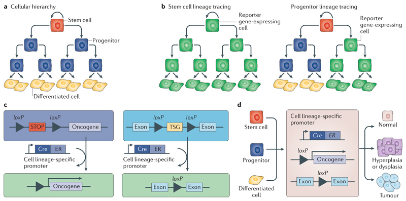
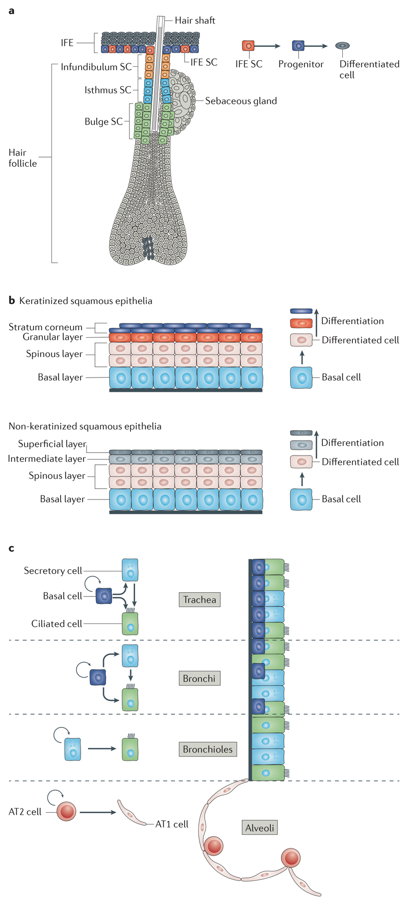
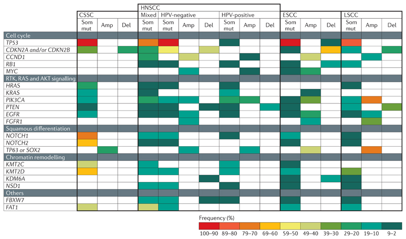
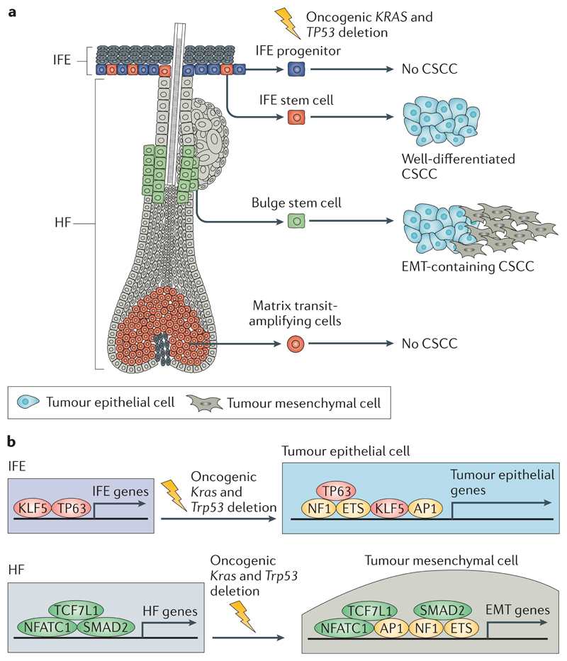
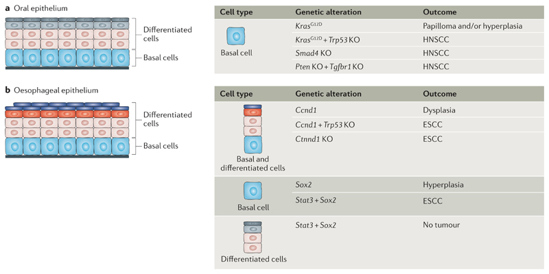
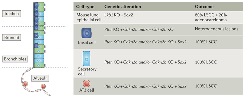
Squamous cell carcinomas (SCCs) are among the most prevalent human cancers. SCC comprises a wide range of tumours originated from diverse anatomical locations that share common genetic mutations and expression of squamous differentiation markers. SCCs arise from squamous and non-squamous epithelial tissues. Here, we discuss the different studies in which the cell of origin of SCCs has been uncovered by expressing oncogenes and/or deleting tumour suppressor genes in the different cell lineages that compose these epithelia. We present evidence showing that the squamous differentiation phenotype of the tumour depends on the type of mutated oncogene and the cell of origin, which dictate the competence of the cells to initiate SCC formation, as well as on the aggressiveness and invasive properties of these tumours.


Conflict of interest statement

Competing interests

The authors declare no competing financial interests.

Figures

Fig 1
Fig 1. Lineage tracing and the cells of origin of cancer.
Many cancers arise from epithelial tissues, which are maintained by stem cells and their progeny. Stem cells are at the top of the cellular hierarchy and have the ability to self-renew and generate progenitors, which can self-renew and give rise to progenitors and terminally differentiated cells (part a). Tamoxifen-induced activation of the Cre recombinase under a cell lineage-specific promoter, for example, a stem cell-specific or progenitor-specific promoter, leads to the elimination of the STOP cassette between loxP sites, resulting in the expression of the reporter gene (part b), the expression of the oncogene or the deletion of the tumour suppressor gene (TSG) between loxP sites (part c) in the respective cell population and its progeny (parts b and c). If a progenitor-specific promoter is used, the reporter gene is expressed in progenitors and their progeny and in differentiated cells but not in the stem cell population. A progenitor usually has a limited lifetime, and thus, the reporter expression may be lost over time (part b, right panel). Conditional activation of oncogenes or deletion of TSGs has allowed the identification of the cells of origin in different mouse tumour models (part d). ER, oestrogen receptor.
Fig 2
Fig 2. Architecture and cellular hierarchy present in the tissues from which SCC arise.
a | The different skin compartments are maintained by their own resident stem cells (SCs). The epidermis is composed of the interfollicular epidermis (IFE), hair follicles and sebaceous glands. The different anatomical regions that form the hair follicle, namely, bulge, infundibulum, isthmus and sebaceous glands, have their own pool of SCs. The hair follicle SCs are slow-cycling cells, residing below the sebaceous gland, in the permanent region of hair follicles. During physiological conditions, hair follicle SCs sustain the cyclic production of the hair, giving rise to transit-amplifying progenitors that rapidly divide and differentiate into the different concentric hair follicles lineages. b | The squamous epithelia of the skin, oral cavity, head and neck and oesophagus are composed of a layer of basal proliferative cells and several suprabasal layers of differentiated cells that progressively flatten before being lost. In keratinized squamous epithelium (for example, oesophageal epithelium), the differentiated cells are enucleated and shed from the surface; these amorphous keratinized ghost cells are called squames. The inner surface of the body is lined with non-keratinized stratified squamous epithelium (for example, oral epithelium), which is characterized by superficial cells that are flattened and nucleated. c | The different lung compartments are maintained by their own pool of SCs during homeostasis. Multipotent basal SCs maintain the mouse trachea and bronchi and give rise to secretory and ciliated cells. In the bronchioles, secretory cells represent a bipotent population of SCs that self-renew and give rise to ciliated cells. In the alveoli, bipotent type 2 (AT2) SCs self-renew and give rise to type 1 (AT1) and AT2 cells.
Fig 3
Fig 3. Common genetic alterations found in the different types of SCC.
Squamous cell carcinomas (SCCs) from different body locations (cutaneous SCC (CSCC), oesophageal SCC (ESCC), head and neck SCC (HNSCC) and lung SCC (LSCC)) have somatic mutations (Som mut), amplifications (Amp) and deletions (Del) in genes controlling the cell cycle, the receptor tyrosine kinase (RTK), RAS and AKT signalling pathways, squamous differentiation and chromatin remodelling. The colour code represents the frequency of a given alteration among patients with each disease subtype. The white fields indicate that Del, Amp or Som mut have not been described for a given gene and SCC type. CCND1, cyclin D1; CDKN2, cyclin-dependent kinase inhibitor 2; EGFR, epidermal growth factor receptor; FAT1, FAT atypical cadherin 1; FBXW7, F-box and WD repeat domain-containing 7; FGFR1, fibroblast growth factor receptor 1; HPV, human papilloma virus; KDM6A, lysine demethylase 6A; KMT2C, lysine methyltransferase 2C; PIK3CA, phosphatidylinositol-4,5-bisphosphate 3-kinase catalytic subunit-α; NSD1, nuclear receptor-binding SET domain protein 1.
Fig 4
Fig 4. The cells at the origin of CSCC.
a | Activation of oncogenic Kras and deletion of Trp53 in interfollicular epidermis (IFE) and hair follicle (HF) stem cells lead to cutaneous squamous cell carcinoma (CSCC) formation, whereas these gene alterations in IFE progenitors or matrix transit-amplifying cells do not lead to CSCC formation. Oncogenic activation of Kras and deletion of Trp53 in IFE stem cells lead to the generation of well-differentiated CSCCs, whereas activation of the same oncogenic hits in HF stem cells leads to the generation of CSCCs with epithelial to mesenchymal transition (EMT) features. b | The transcriptional and epigenetic landscape of the cell of origin influences tumour differentiation. Upon oncogenic Kras expression and Trp53 deletion, a core of transcription factors (including members of the adaptor protein 1 (AP1), E26 transformation-specific (ETS) and nuclear factor 1 (NF1) families) promote tumour gene expression independently of the cell of origin. In addition to this core of transcription factors, lineage-specific transcription factors controlled by the cells of origin of CSCCs influence the specific differentiation of the tumours. Tumour protein 63 (TP63) and Krüppel-like factor 5 (KLF5) promote the expression of IFE genes and the development of well-differentiated squamous cell carcinomas (SCCs), whereas SMAD family member 2 (SMAD2), nuclear factor of activated T cell, cytoplasmic 1 (NFATC1) and transcription factor 7-like 1 (TCF7L1) promote the expression of HF genes and the development of SCCs in which EMT occurs.
Fig 5
Fig 5. The cells of origin in HNSCC and ESCC.
a | The cells at the origin of head and neck squamous cell carcinoma (HNSCC) are shown. Basal cells of the oral epithelia can give rise to hyperplasia and/or papilloma formation upon oncogenic Kras activation. HNSCC can result from the activation of Kras, the combination of activation of Kras and deletion of Trp53, SMAD family member 4 (Smad4) deletion or double deletion of Pten and transforming growth factorbeta receptor type-1 (Tgfbr1) in basal cells. b | The cells at the origin of oesophageal squamous cell carcinoma (ESCC) are shown. Activation of cyclin D1 (Ccnd1) in combination with Trp53 deletion and deletion of catenin delta-1 (Ctnnd1) in oesophageal epithelial cells leads to ESCC formation. Expression of signal transducer and activator of transcription 3 (Stat3) and SRY-box 2 (Sox2) in basal cells but not in suprabasal cells promotes ESCC. KO, knockout.
Fig 6
Fig 6. SOX2 promotes LSCC differentiation irrespective of the cell of origin.
Transcription factor SRY-box2 (SOX2) promotes squamous cell fate in lung tumours regardless of the cell of origin. Sox2 overexpression and serine/threonine kinase 11 (Lkb1 deletion in cells of the lung epithelium lead to lung squamous cell carcinoma (LSCC) and adenocarcinoma. In the absence of Sox2 overexpression, Pten and cyclin-dependent kinase inhibitor 2A (Cdkn2a) and/or Cdkn2b deletion in basal cells leads to heterogeneous lesions including adenocarcinoma and LSCCs. Sox2 overexpression and Pten and Cdkn2a and/or Cdkn2b deletion in basal, secretory or type 2 (AT2) cells lead to the formation of LSCCs. KO, knockout.

References

    1. Dotto GP, Rustgi AK. Squamous cell cancers: a unified perspective on biology and genetics. Cancer Cell. 2016;29:622–637. [This is a landmark review summarizing the aetiology and genetic determinants of SCCs arising from different body locations.] - PMC - PubMed
    1. Alam M, Ratner D. Cutaneous squamous-cell carcinoma. N Engl J Med. 2001;344:975–983. - PubMed
    1. Herbst RS, Heymach JV, Lippman SM. Lung cancer. N Engl J Med. 2008;359:1367–1380. - PMC - PubMed
    1. Leemans CR, Braakhuis BJ, Brakenhoff RH. The molecular biology of head and neck cancer. Nat Rev Cancer. 2011;11:9–22. - PubMed
    1. Rustgi AK, El-Serag HB. Esophageal carcinoma. N Engl J Med. 2014;371:2499–2509. - PubMed

Publication types

MeSH terms