Modulation of Corticospinal Excitability Depends on the Pattern of Mechanical Tactile Stimulation
- PMID: 29849557
- PMCID: PMC5903327
- DOI: 10.1155/2018/5383514
Modulation of Corticospinal Excitability Depends on the Pattern of Mechanical Tactile Stimulation
Abstract
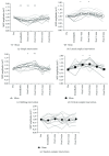
We investigated the effects of different patterns of mechanical tactile stimulation (MS) on corticospinal excitability by measuring the motor-evoked potential (MEP). This was a single-blind study that included nineteen healthy subjects. MS was applied for 20 min to the right index finger. MS intervention was defined as simple, lateral, rubbing, vertical, or random. Simple intervention stimulated the entire finger pad at the same time. Lateral intervention stimulated with moving between left and right on the finger pad. Rubbing intervention stimulated with moving the stimulus probe, fixed by protrusion pins. Vertical intervention stimulated with moving in the forward and backward directions on the finger pad. Random intervention stimulated to finger pad with either row protrudes. MEPs were measured in the first dorsal interosseous muscle to transcranial magnetic stimulation of the left motor cortex before, immediately after, and 5-20 min after intervention. Following simple intervention, MEP amplitudes were significantly smaller than preintervention, indicating depression of corticospinal excitability. Following lateral, rubbing, and vertical intervention, MEP amplitudes were significantly larger than preintervention, indicating facilitation of corticospinal excitability. The modulation of corticospinal excitability depends on MS patterns. These results contribute to knowledge regarding the use of MS as a neurorehabilitation tool to neurological disorder.
Figures


Similar articles
-
The effects of mechanical tactile stimulation on corticospinal excitability and motor function depend on pin protrusion patterns.Sci Rep. 2019 Nov 13;9(1):16677. doi: 10.1038/s41598-019-53275-2. Sci Rep. 2019. PMID: 31723202 Free PMC article.
-
Corticospinal Excitability During Actual and Imaginary Motor Tasks of Varied Difficulty.Neuroscience. 2018 Nov 1;391:81-90. doi: 10.1016/j.neuroscience.2018.08.011. Epub 2018 Aug 19. Neuroscience. 2018. PMID: 30134204
-
Whole-hand water flow stimulation increases motor cortical excitability: a study of transcranial magnetic stimulation and movement-related cortical potentials.J Neurophysiol. 2015 Feb 1;113(3):822-33. doi: 10.1152/jn.00161.2014. Epub 2014 Nov 5. J Neurophysiol. 2015. PMID: 25376780
-
Tactile Perception of Right Middle Fingertip Suppresses Excitability of Motor Cortex Supplying Right First Dorsal Interosseous Muscle.Neuroscience. 2022 Jul 1;494:82-93. doi: 10.1016/j.neuroscience.2022.05.012. Epub 2022 May 16. Neuroscience. 2022. PMID: 35588919
-
Changes in corticospinal excitability during motor imagery by physical practice of a force production task: Effect of the rate of force development during practice.Neuropsychologia. 2024 Aug 13;201:108937. doi: 10.1016/j.neuropsychologia.2024.108937. Epub 2024 Jun 10. Neuropsychologia. 2024. PMID: 38866222
Cited by
-
Effectiveness and functional magnetic resonance imaging outcomes of Tuina therapy in patients with post-stroke depression: A randomized controlled trial.Front Psychiatry. 2022 Jun 30;13:923721. doi: 10.3389/fpsyt.2022.923721. eCollection 2022. Front Psychiatry. 2022. PMID: 35845459 Free PMC article.
-
Discrimination of the moving direction is improved depending on the pattern of the mechanical tactile stimulation intervention.BMC Neurosci. 2024 Mar 5;25(1):15. doi: 10.1186/s12868-024-00855-2. BMC Neurosci. 2024. PMID: 38443782 Free PMC article.
-
The Repetitive Mechanical Tactile Stimulus Intervention Effects Depend on Input Methods.Front Neurosci. 2020 Apr 28;14:393. doi: 10.3389/fnins.2020.00393. eCollection 2020. Front Neurosci. 2020. PMID: 32410954 Free PMC article.
-
Balance in Blind Subjects: Cane and Fingertip Touch Induce Similar Extent and Promptness of Stance Stabilization.Front Neurosci. 2018 Sep 11;12:639. doi: 10.3389/fnins.2018.00639. eCollection 2018. Front Neurosci. 2018. PMID: 30254565 Free PMC article.
-
Effects of repetitive mechanical tactile stimulation interventions with stationary and moving patterns on paired-pulse depression.BMC Neurosci. 2025 Jul 26;26(1):46. doi: 10.1186/s12868-025-00960-w. BMC Neurosci. 2025. PMID: 40713502 Free PMC article.
References
Publication types
MeSH terms
LinkOut - more resources
Full Text Sources
Other Literature Sources

