. 2018 Mar 23;9(36):24272-24282.
doi: 10.18632/oncotarget.24917. eCollection 2018 May 11.

Macrophage inflammatory factors promote epithelial-mesenchymal transition in breast cancer

Affiliations

Macrophage inflammatory factors promote epithelial-mesenchymal transition in breast cancer

Robert B Bednarczyk et al. Oncotarget. .

Abstract

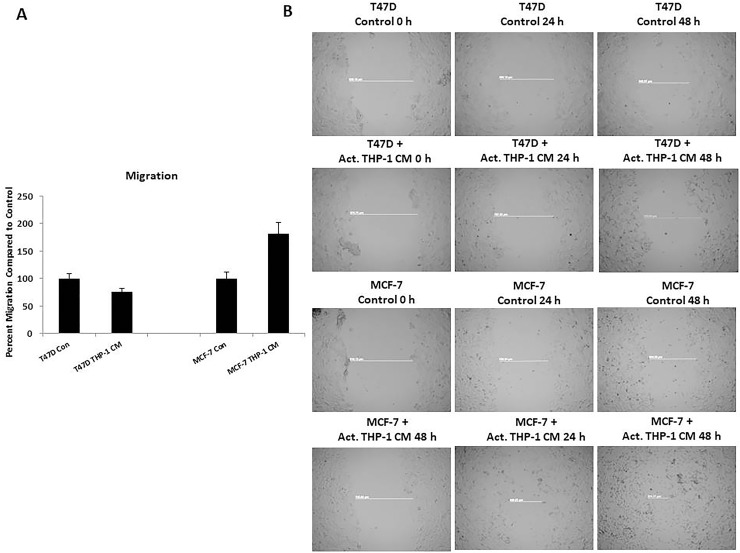
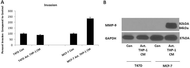
The majority of breast cancers (90-95%) arise due to mediators distinct from inherited genetic mutations. One major mediator of breast cancer involves chronic inflammation. M1 macrophages are an integral component of chronic inflammation and the breast cancer tumor microenvironment (TME). Previous studies have demonstrated that up to 50% of the breast tumor comprise of tumor-associated macrophages (TAMs) and increased TAM infiltration has been associated with poor patient prognosis. Furthermore, breast cancer associated deaths are predominantly attributed to invasive cancers and metastasis with epithelial-mesenchymal transition (EMT) being implicated. In this study, we investigated the effects of cellular crosstalk between TAMs and breast cancer using an in vitro model system. M1 polarized THP-1 macrophage conditioned media (CM) was generated and used to evaluate cellular and functional changes of breast cancer lines T47D and MCF-7. We observed that T47D and MCF-7 exhibited a partial EMT phenotype in the presence of activated THP-1 CM. Additionally, MCF-7 displayed a significant increase in migratory and invasive properties. We conclude that M1 secretory factors can promote a partial EMT of epithelial-like breast cancer cells. The targeting of M1 macrophages or their secretory components may inhibit EMT and limit the invasive potential of breast cancer.

Keywords: M1 macrophages; epithelial-mesenchymal transition; inflammation; metastasis; secretory factors.


Conflict of interest statement

CONFLICTS OF INTEREST No potential conflicts of interest were disclosed.

Figures

Figure 1
Figure 1. THP-1 cells exhibit an M1 macrophage phenotype following activation with TPA
(A) THP-1 cells express monocyte/macrophage lineage marker CD68. (B) Upon activation with TPA, THP-1 cells express M1 macrophage markers iNOS and IL-6. Unactivated and activated THP-1 cells were seeded overnight and probed with CD68, iNOS, or IL-6 primary antibodies. Fluorescent secondary antibodies were used for immunofluorescence. Pictures were taken using Axiovision Rel 4.8 at 100x magnification. (C) Activated THP-1 cells express pro-inflammatory cytokines including: TNF-α, IL-1β, and IL-8. A human inflammation array was performed on unactivated or activated THP-1 CM (48 h). Cytokine/chemokine blots were analyzed using ImageJ and optical density determined and normalized ± SEM of two independent experiments.
Figure 2
Figure 2. T47D and MCF-7 cells exhibited spindle-like morphology, decreased E-cadherin, and decreased viability viable cells following treatment with activated THP-1 CM
(A) Treatment of T47D and MCF-7 with activated THP-1 CM promotes mesenchymal-like morphological changes. Breast cancer cells were treated with unactivated or activated THP-1 CM for 24 h and cell morphology was observed by phase contrast microscopy. Pictures were taken at 20x magnification using phase contrast microscopy. (B) T47D and MCF-7 were treated for 24 h with control (5% CS-RPMI) or activated THP-1 CM and probed with E-cadherin primary antibodies. Fluorescent secondary antibodies in addition to DAPI (nuclear stain) were used for immunofluorescence. Pictures were taken using Axiovision Rel 4.8 at 100x magnification. (C) Activated THP-1 CM leads to decreased number of viable T47D and MCF-7 cells at 24 and 48 h. A trypan blue exclusion assay was performed to evaluate the number of viable cells. Data represents the number of viable cells in duplicates ± SEM of two independent experiments.
Figure 3
Figure 3. Spindle-like morphology and reduced viability is observed following co-culture with activated THP-1 cells
(A) Co-culture of activated THP-1 cells with either T47D or MCF-7 promotes mesenchymal-like morphology. Breast cancer cells were co-cultured for 24 h and cell morphology was observed. Pictures were taken at 10x magnification using phase contrast microscopy. (B) A decreased number of viable breast cancer cells was observed following co-culture with activated THP-1 cells for 24 h. A trypan blue exclusion assay was performed to evaluate viability. Data represents the number of viable cells in duplicates ± SEM of two independent experiments in duplicates.
Figure 4
Figure 4. T47D and MCF-7 cells exhibit increased nuclear localization of EMT transcription factors
(A) Increased nuclear localization of NF-κB and Snail is observed in T47D. (B) MCF-7 displays increased nuclear localization of NF-κB, Snail, and Slug. (C) MDA-MB 231 robustly expresses mesenchymal markers Vimentin and Slug. T47D, MCF-7, and MDA-MB 231 were treated with activated THP-1 CM for 6 h, 12 h, 24 h, and 48 h. Nuclear extractions were prepared and Western blots performed probing for: NF-κB (65 kDa), Slug (30 kDa), Snail (29 kDa), Vimentin (57 kDa), β-Actin (45 kDa), and Lamin (63, 74 kDa). Relative density of NF-κB, Snail, and Slug is shown normalized to Lamin loading controls ± SEM of two independent experiments.
Figure 5
Figure 5. THP-1 CM promotes MCF-7 migration
(A) MCF-7 exhibits increased migration at 18 h in the presence of activated THP-1 CM. Breast cancer migration was evaluated using Boyden chamber assays. Pictures were taken using brightfield microscopy at 5x magnification counting four fields per insert. Data is represented as percent migration compared to control in duplicates ± SEM of two independent experiments. (B) Scratch wound assay confirms increased MCF-7 migration. Scratch-wound assay was performed on T47D and MCF-7 treated with control (5% CS-RPMI) or activated THP-1 CM with mitomycin C (2 μg/mL) for 24 h. Pictures were taken at 0 h, 24 h, and 48 h following control or CM treatments using phase contrast microscopy and Axiovision Rel 4.8 at 5x magnification.
Figure 6
Figure 6. Activated THP-1 CM treatment enhances MCF-7 invasion and MMP-9 expression
(A) MCF-7 exhibits increased invasion at 18 h in the presence of activated THP-1 CM. Breast cancer invasion was evaluated using Boyden chamber assays. Pictures were taken using brightfield microscopy at 5x magnification counting four fields per insert. Data is represented as percent invasion compared to control in duplicates ± SEM of two independent experiments. (B) There is increased expression of MMP-9 by MCF-7 with 24 h activated THP-1 CM treatment. MMP-9 (84, 92 kDa) and GAPDH (37 kDa) expression was evaluated using Western blots.

References

    1. Siegel RL, Miller KD, Jemal A. Cancer statistics, 2018. CA Cancer J Clin. 2016;68:7–30. - PubMed
    1. DeSantis CE, Fedewa SA, Goding Sauer A, Kramer JL, Smith RA, Jemal A. Breast cancer statistics, 2015: Convergence of incidence rates between black and white women. CA Cancer J Clin. 2016;66:31–42. - PubMed
    1. Weigelt B, Peterse JL, van 't Veer LJ. Breast cancer metastasis: Markers and models. Nat Rev Cancer. 2005;5:591–602. - PubMed
    1. Dossus L, Benusiglio PR. Lobular breast cancer: Incidence and genetic and non-genetic risk factors. Breast Cancer Res. 2015;17:37. - PMC - PubMed
    1. Krieger N, Chen JT, Waterman PD. Decline in US breast cancer rates after the women's health initiative: Socioeconomic and racial/ethnic differentials. Am J Public Health. 2010;100:972. - PMC - PubMed

LinkOut - more resources