Case Reports
. 2018 Apr;10(4):E289-E295.
doi: 10.21037/jtd.2018.03.77.

Primary aortic sarcoma in arch and descending aorta: a case report and literature review

Affiliations
Case Reports

Primary aortic sarcoma in arch and descending aorta: a case report and literature review

Zhi-Yuan Wu et al. J Thorac Dis. 2018 Apr.

Abstract

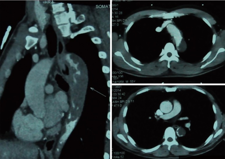
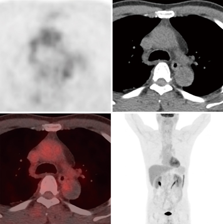
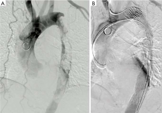
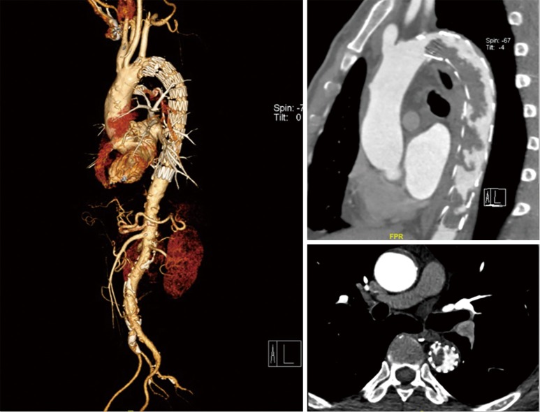
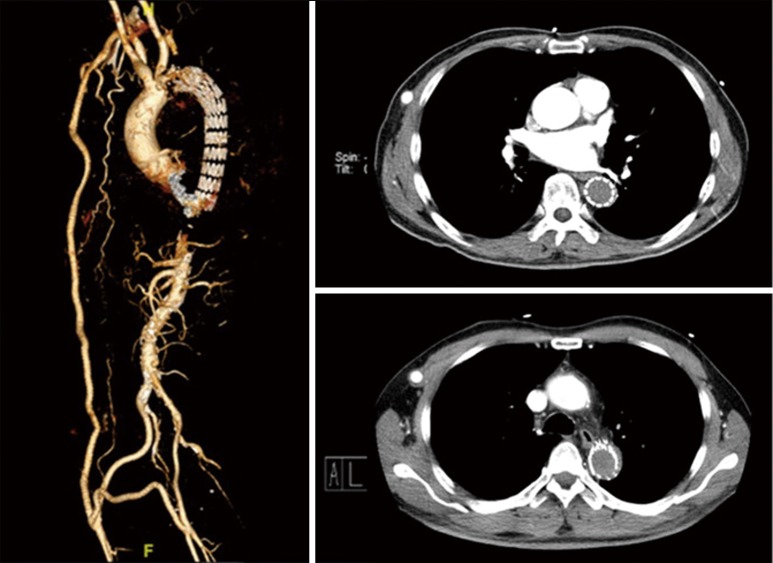
Aortic sarcoma is a rare entity. In this study, we report a case of a 56-year-old man with complaints of pain and numbness of bilateral lower extremities. An endovascular aortic repair was finally adopted to prevent recurrent embolic events. An endo-biopsy was performed and showed aortic sarcoma. Axillary bifemoral and femoro-femoral cross-over bypass surgeries were taken to supply blood to the lower extremities in the 6th month after the first operation. He finally passed away in the 37th month. Aortic sarcoma should be taken into consideration as one of the possible etiologies for massive thrombus in aorta. Palliative surgeries such as bypass, endovascular aortic repair can also be an alternative to treat aortic sarcoma.

Keywords: Aorta; bypass; endovascular aortic repair; sarcoma.


Conflict of interest statement

Conflict of Interest: The authors have no conflicts of interest to declare.

Figures

Figure 1
Figure 1
Atherosclerosis and multiple ulcerating plaques with thrombus in aorta.
Figure 2
Figure 2
PET/CT revealed no signs of increased intake in the lesion, with the SUVmax value of 2.3. PET/CT, positron emission tomography/computed tomography.
Figure 3
Figure 3
A thoracic endovascular aortic repair (TEVAR) was performed for this patient. (A) Angiography showed the filling-defect part in the aorta; (B) completion angiography demonstrated the blood flow of thoracic aorta and branches were patent.
Figure 4
Figure 4
Aorta CTA showed thrombus in the lumen of stent covered in arch of aorta and descending aorta in the 6th month after first endovascular management. CTA, computed tomography angiography.
Figure 5
Figure 5
A CTA showed patent in the bridge vessel in the 22nd month. CTA, computed tomography angiography.

References

    1. Salm R. Primary fibrosarcoma of aorta. Cancer 1972;29:73-83. 10.1002/1097-0142(197201)29:1<73::AID-CNCR2820290113>3.0.CO;2-V - DOI - PubMed
    1. Wright EP, Glick AD, Virmani R, et al. Aortic intimal sarcoma with embolic metastases. Am J Surg Pathol 1985;9:890-7. 10.1097/00000478-198512000-00005 - DOI - PubMed
    1. Thalheimer A, Fein M, Geissinger E, et al. Intimal angiosarcoma of the aorta: report of a case and review of the literature. J Vasc Surg 2004;40:548-53. 10.1016/j.jvs.2004.06.035 - DOI - PubMed
    1. Nishida N, Yutani C, Ishibashi-Ueda H, et al. Histopathological characterization of aortic intimal sarcoma with multiple tumor emboli. Pathol Int 2000;50:923-7. 10.1046/j.1440-1827.2000.01144.x - DOI - PubMed
    1. Chiche L, Mongredien B, Brocheriou I, et al. Primary tumors of the thoracoabdominal aorta: surgical treatment of 5 patients and review of the literature. Ann Vasc Surg 2003;17:354-64. 10.1007/s10016-003-0018-x - DOI - PubMed

Publication types

LinkOut - more resources