

Successful phased approach to a patient with synchronous traumatic descending aortic pseudoaneurysm and bronchial rupture
- PMID: 29850175
- PMCID: PMC5949447
- DOI: 10.21037/jtd.2018.04.51
Successful phased approach to a patient with synchronous traumatic descending aortic pseudoaneurysm and bronchial rupture
Abstract
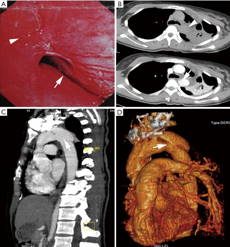
A 34-year-old woman was referred to our center because of collapsed left lung and left main bronchial stenosis 1 week after a vehicular accident. Bronchoscopic observation revealed stenosis in the left main and lobar bronchus. Computed tomography (CT) angiography found traumatic descending aortic pseudoaneurysm after admission. Phased intervention strategy was adopted. The aortic rupture was repaired with endovascular stent firstly, followed by sleeve reconstruction of the left main bronchus through posterolateral thoracotomy 11 days later. She recovered uneventfully and resulted in an excellent long-term outcome. The diagnosis and treatment of this case is discussed.
Keywords: Aortic pseudoaneurysm; blunt chest trauma; bronchial rupture.
Conflict of interest statement
Conflicts of Interest: The authors have no conflicts of interest to declare.
Figures

References
-
- Exadaktylos AK, Duwe J, Eckstein F, et al. The role of contrast-enhanced spiral CT imaging versus chest X-rays in surgical therapeutic concepts and thoracic aortic injury: a 29-year Swiss retrospective analysis of aortic surgery. Cardiovasc J S Afr 2005;16:162-5. - PubMed
Publication types
LinkOut - more resources
Full Text Sources
Other Literature Sources