Case Reports
. 2018 Mar 28;10(3):e2381.
doi: 10.7759/cureus.2381.

Surgical Reconstruction to Allow Endovascular Access for Flow Diversion of Giant Cavernous Aneurysm: A Combined Approach

Affiliations
Case Reports

Surgical Reconstruction to Allow Endovascular Access for Flow Diversion of Giant Cavernous Aneurysm: A Combined Approach

Visish M Srinivasan et al. Cureus. .

Abstract

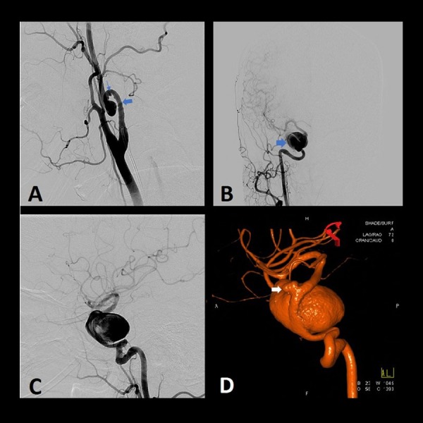
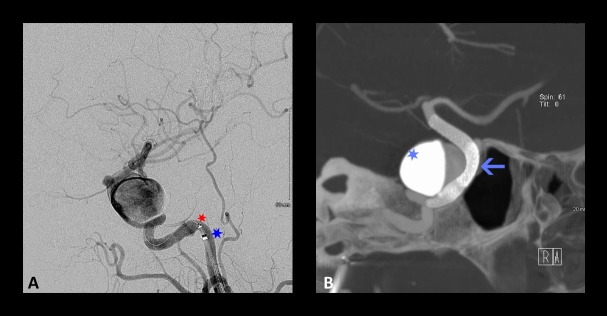
Giant cavernous aneurysms of the internal carotid artery (ICA) are challenging lesions associated with high surgical morbidity. Prior to the past several years, these were treated by surgical reconstruction, proximal ligation, or stent-assisted coiling techniques. Flow diversion has become the standard of care for these lesions, providing a high rate of obliteration with a much better safety profile. However, flow diverters rely upon a navigable vasculature and, usually, a tri-axial support system. Cases in which such access is difficult require unique approaches to combine the strengths of both surgical and endovascular therapy. A woman with a giant cavernous ICA aneurysm and an ophthalmic artery aneurysm presented for treatment, but access was challenging due to cervical ICA tortuosity and pseudoaneurysms. We elected a staged, combined approach with surgical reconstruction of the cervical ICA followed by flow diverter placement for the intracranial aneurysms. Our case features an "outside-the-box" approach that synergistically applied both microsurgical and endovascular techniques to treat a challenging pathology. Classic microsurgical techniques remain important in cases that are refractory or not amenable to endovascular therapy alone.

Keywords: carotid endarterectomy; cerebral aneurysm; cervical aneurysm; flow diversion; giant aneurysm; hunterian ligation.


Conflict of interest statement

Dr. Kan is a consultant for Medtronic and Styker.

Figures

Figure 1
Figure 1. Preoperative angiography of the right common carotid artery showing cervical and intracranial aneurysms
Preoperative angiography of the right common carotid artery showing cervical and intracranial aneurysms. (A) Lateral – cervical, (B) anteroposterior – head, (C) Lateral – head, (D) three-dimensional (3D) reconstruction. The cervical loop (large arrow) and pseudoaneurysms (thin arrows) precluded endovascular access to the giant cavernous (large blue arrow) and smaller ophthalmic aneurysm (white arrow).
Figure 2
Figure 2. Comparison of preoperative and postoperative angiograms of the right cervical internal carotid artery
Comparison of preoperative and postoperative angiograms of the right cervical internal carotid artery. (A) Three-dimensional (3D) angiogram showing a 360-degree loop with three pseudoaneurysms. (B) Postoperative right common carotid artery angiogram, showing patent reanastomosis and favorable, straight cervical segment for endovascular access.
Figure 3
Figure 3. Intraoperative photograph of right cervical exposure
The external carotid artery (ECA), common carotid artery (CCA), internal carotid artery (ICA), and excised pseudoaneurysm-containing loop are labeled. Note that the exposure extends more rostally than that used for a typical carotid endarterectomy.
Figure 4
Figure 4. Postoperative angiography via right common carotid artery two weeks after surgical reconstruction
Postoperative angiography via right common carotid artery two weeks after surgical reconstruction. Partial thrombosis (Hunterian ligation phenomenon) of the giant cavernous aneurysm following temporary trapping for the surgical resection of the carotid loop is seen in (A) anteroposterior view, (B) lateral view, and (C) three-dimensional (3D) reconstruction. Arrows indicate the scalloping of the aneurysm, with contrast opacifying the patent portion of the lumen, whereas the thrombus is not visible.
Figure 5
Figure 5. Intraprocedural angiography for the placement of a flow diverter
Intraprocedural angiography for the placement of a flow diverter. (A) Lateral angiography showing the positioning of the intermediate catheter (blue star) and the microcatheter (red star) as part of a tri-axial catheter system and (B) Cone-beam computed tomography (CBCT) (Dyna CT) showing the positioning of two partially overlapping devices (arrow) between the distal petrous segment and the communicating segment of the ICA. Contrast stagnation can be appreciated within the aneurysm (light blue star).
Figure 6
Figure 6. Postoperative angiography via right common carotid artery
Postoperative angiography via right common carotid artery showing the obliteration of both intracranial aneurysms in (A) anteroposterior and (B) lateral view. The flow diverters span the distal petrous segment to the communicating segment of the internal carotid artery (ICA).

References

    1. The expanding realm of endovascular neurosurgery: flow diversion for cerebral aneurysm management. Krishna C, Sonig A, Natarajan SK, Siddiqui AH. https://www.ncbi.nlm.nih.gov/pubmed/25624975. Methodist Debakey Cardiovasc J. 2014;10:214–219. - PMC - PubMed
    1. Earlier times in aneurysm surgery. Drake CG. Clin Neurosurg. 1985;32:41–50. - PubMed
    1. Cerebrovascular neurosurgery in evolution: the endovascular paradigm. Sorkin GC, Dumont TM, Eller JL, et al. Neurosurgery. 2014;74:191–197. - PubMed
    1. Reconstruction of the ligated external carotid artery for embolization of cervicofacial arteriovenous malformations. Riles TS, Berenstein A, Fisher FS, Persky MS, Madrid M. J Vasc Surg. 1993;17:491–498. - PubMed
    1. The combined approach to intracranial aneurysm treatment. Alexander BL, Riina HA. Surg Neurol. 2009;72:596–606. - PubMed

Publication types

LinkOut - more resources