

Transcatheter Arterial Embolization Therapy for Huge Renal Cysts: Two Case Reports
- PMID: 29850462
- PMCID: PMC5968287
- DOI: 10.1159/000489088
Transcatheter Arterial Embolization Therapy for Huge Renal Cysts: Two Case Reports
Abstract
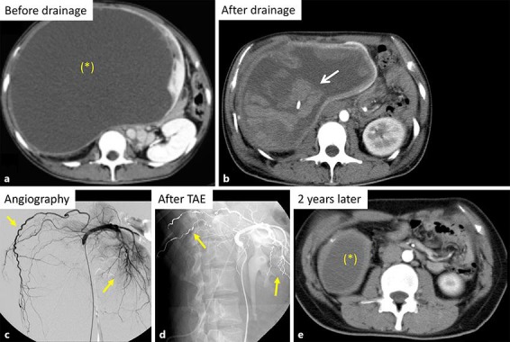
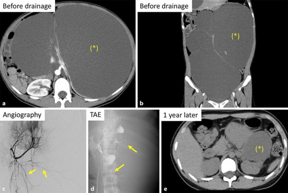
We encountered 2 patients with symptomatic huge simple renal cysts. In case 1, 4,000 mL of cyst fluid was drained via a catheter, but intracystic bleeding occurred immediately afterwards. Transcatheter arterial embolization (TAE) was performed, after which the bleeding stopped, and cyst drainage was repeated successfully. After 2 years, the total cyst volume was reduced from 11,775 mL to 75.4 mL. In case 2, TAE was performed prophylactically before drainage. Subsequently, 9,400 mL of fluid was removed from multiple cysts. After 1 year, the total cyst volume was reduced from 9,215 mL to 633 mL without bleeding. Based on these 2 cases, prophylactic TAE before drainage may be useful in patients with huge renal cysts.
Keywords: Cyst drainage; Simple renal cyst; Transcatheter arterial embolization.
Figures
Similar articles
-
Nonparasitic solitary huge liver cysts causing intracystic hemorrhage or obstructive jaundice.J Hepatobiliary Pancreat Surg. 2002;9(6):764-8. doi: 10.1007/s005340200107. J Hepatobiliary Pancreat Surg. 2002. PMID: 12658414
-
Treatment of symptomatic polycystic liver disease: transcatheter super-selective hepatic arterial embolization using a mixture of NBCA and iodized oil.Abdom Imaging. 2013 Jun;38(3):465-73. doi: 10.1007/s00261-012-9931-1. Abdom Imaging. 2013. PMID: 22743841
-
Percutaneous transcatheter hepatic artery embolization for liver cysts in autosomal dominant polycystic kidney disease.Am J Kidney Dis. 2007 Jun;49(6):744-52. doi: 10.1053/j.ajkd.2007.03.018. Am J Kidney Dis. 2007. PMID: 17533017
-
[Transcatheter embolization of huge renal arteriovenous aneurysm: a case report--review of 270 cases of renal arteriovenous fistula reported in Japan].Hinyokika Kiyo. 1991 Mar;37(3):273-7. Hinyokika Kiyo. 1991. PMID: 2069108 Review. Japanese.
-
[Transcatheter arterial embolization in patients with renal arteriovenous malformation: a report of two cases].Hinyokika Kiyo. 1989 Oct;35(10):1761-5. Hinyokika Kiyo. 1989. PMID: 2692440 Review. Japanese.
Cited by
-
Transcatheter arterial embolization in a haemorrhagic renal cyst with "rising tide sign".Clin Exp Emerg Med. 2022 Dec;9(4):377-379. doi: 10.15441/ceem.22.190. Epub 2022 Jul 8. Clin Exp Emerg Med. 2022. PMID: 35793789 Free PMC article. No abstract available.
-
Transcatheter arterial embolisation of a haemorrhagic renal cyst.BMJ Case Rep. 2020 Jul 1;13(7):e236628. doi: 10.1136/bcr-2020-236628. BMJ Case Rep. 2020. PMID: 32611658 Free PMC article. No abstract available.
References
-
- Agarwal MM, Hemal AK. Surgical management of renal cystic disease. Curr Urol Rep. 2011 Feb;12((1)):3–10. - PubMed
-
- Suwabe T, Ubara Y, Sumida K, Hayami N, Hiramatsu R, Yamanouchi M, et al. Clinical features of cyst infection and hemorrhage in ADPKD: new diagnostic criteria. Clin Exp Nephrol. 2012 Dec;16((6)):892–902. - PubMed
-
- Ubara Y, Tagami T, Sawa N, Katori H, Yokota M, Takemoto F, et al. Renal contraction therapy for enlarged polycystic kidneys by transcatheter arterial embolization in hemodialysis patients. Am J Kidney Dis. 2002 Mar;39((3)):571–579. - PubMed
Publication types
LinkOut - more resources
Full Text Sources
Other Literature Sources
