Review
. 2018 Apr 5:2018:5065190.
doi: 10.1155/2018/5065190. eCollection 2018.

Drosophila melanogaster Models of Friedreich's Ataxia

Affiliations
Review

Drosophila melanogaster Models of Friedreich's Ataxia

P Calap-Quintana et al. Biomed Res Int. .

Abstract

Friedreich's ataxia (FRDA) is a rare inherited recessive disorder affecting the central and peripheral nervous systems and other extraneural organs such as the heart and pancreas. This incapacitating condition usually manifests in childhood or adolescence, exhibits an irreversible progression that confines the patient to a wheelchair, and leads to early death. FRDA is caused by a reduced level of the nuclear-encoded mitochondrial protein frataxin due to an abnormal GAA triplet repeat expansion in the first intron of the human FXN gene. FXN is evolutionarily conserved, with orthologs in essentially all eukaryotes and some prokaryotes, leading to the development of experimental models of this disease in different organisms. These FRDA models have contributed substantially to our current knowledge of frataxin function and the pathogenesis of the disease, as well as to explorations of suitable treatments. Drosophila melanogaster, an organism that is easy to manipulate genetically, has also become important in FRDA research. This review describes the substantial contribution of Drosophila to FRDA research since the characterization of the fly frataxin ortholog more than 15 years ago. Fly models have provided a comprehensive characterization of the defects associated with frataxin deficiency and have revealed genetic modifiers of disease phenotypes. In addition, these models are now being used in the search for potential therapeutic compounds for the treatment of this severe and still incurable disease.


Figures

Figure 1
Figure 1
Molecular phylogenetic analysis of frataxin sequences from different species. The picture of Thomas Hunt Morgan was chosen to represent Homo sapiens because, as a result of his work, D. melanogaster became a major model organism in genetics. Methods: evolutionary history was inferred with the maximum likelihood method based on Le and Gascuel model [9]. The tree with the highest log likelihood (−2026.7976) is shown. Initial trees for the heuristic search were obtained automatically by applying the Neighbor-Joining and BioNJ algorithms to a matrix of pairwise distances estimated using a JTT model and then selecting the topology with the superior log likelihood value. A discrete gamma distribution was used to model evolutionary rate differences among sites (5 categories (+G, parameter = 2.4842)). The tree is drawn to scale, with branch lengths representing the number of substitutions per site. The analysis involved 16 amino acid sequences. All positions containing gaps and missing data were eliminated. A total of 90 positions were present in the final dataset. Evolutionary analyses were conducted in MEGA7 [10].
Figure 2
Figure 2
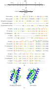
The Drosophila frataxin ortholog. (a) Genomic organization of the human (FXN) and the fly (fh) genes encoding frataxin. FXN is located in 9q21.11 and contains seven exons. fh is located in chromosome X: 8C14 and has two exons. (b) Multiple alignment of the frataxin protein sequences of Homo sapiens, Mus musculus, D. melanogaster, Caenorhabditis elegans, and Saccharomyces cerevisiae. The letters indicate the amino acid in each position, and the colors classify the amino acids according to their biochemical properties, as described in the MEGA7 program [10]. Invariant amino acids are marked with an asterisk. (c) The 3D structure prediction of the frataxin protein using the Phyre 2 [11] and Chimera 1.12 software [12]; α-helixes appear in blue and β-sheets in green.
Figure 3
Figure 3
The GAL4/UAS system, adapted from yeast, involves the use of two transgenic lines in Drosophila [13]. One line carries the GAL4 transcription factor under the control of a promoter of known expression pattern (the driver line), and the other line contains the transgene of interest downstream of UAS (the responder line). Many GAL4 driver lines are available, carrying the promoters of genes such as actin (ubiquitous), elav (pan-neuronal), repo (glial cells), neur (sensory organs), and GMR (eye). This system is very versatile and allows the expression of specific genes or gene constructs to be induced or suppressed. Triangles indicate a wild-type or mutant protein; the hairpins represent double-stranded RNA molecules that mediate RNAi.
Figure 4
Figure 4
Schematic design of a genetic (a) or chemical (b) screen to identify genetic modifiers or potential therapeutic compounds in FRDA using Drosophila as a model organism. The effect of a genetic modifier or drug is evaluated by monitoring the lifespan and climbing ability of FRDA flies. (c) A UAS-GFP construct is included in this strategy as an internal control to determine whether the drug can interfere with the GAL4/UAS system and the potential dilution of the GAL4 protein due to the presence of two UAS construct. In parallel, the effect of the modifier or drug treatment is analyzed in control flies to identify frataxin interactors. GFP: green fluorescent protein. Vehicle: DMSO/H2O depending on the drug solubility.

References

    1. Harding A. Clinical features and classification of inherited ataxias. Advances in Neurology. 1993;61:1–14. - PubMed
    1. Bidichandani S. I., Delatycki M. B. Friedreich Ataxia. GeneReviews®. 2017.
    1. Campuzano V., Montermini L., Moltò M. D., et al. Friedreich's ataxia: autosomal recessive disease caused by an intronic GAA triplet repeat expansion. Science. 1996;271(5254):1423–1427. doi: 10.1126/science.271.5254.1423. - DOI - PubMed
    1. Koutnikova H., Campuzano V., Foury F., Dollé P., Cazzalini O., Koenig M. Studies of human, mouse and yeast homologues indicate a mitochondrial function for frataxin. Nature Genetics. 1997;16(4):345–351. doi: 10.1038/ng0897-345. - DOI - PubMed
    1. Koutnikova H., Campuzano V., Koenig M. Maturation of wild-type and mutated frataxin by the mitochondrial processing peptidase. Human Molecular Genetics. 1998;7(9):1485–1489. doi: 10.1093/hmg/7.9.1485. - DOI - PubMed

Substances

LinkOut - more resources