

Influence of brightness and contrast adjustments on the diagnosis of proximal caries lesions
- PMID: 29851369
- PMCID: PMC6326395
- DOI: 10.1259/dmfr.20180100
Influence of brightness and contrast adjustments on the diagnosis of proximal caries lesions
Abstract
Objectives: To assess the influence of brightness and contrast adjustments of digital radiographs on the diagnosis of proximal caries lesions, and to compare with observers' preferences for subjective image quality.
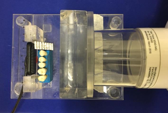
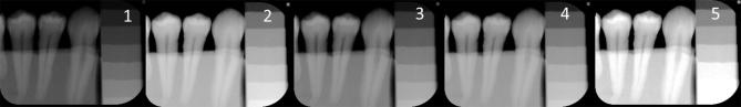
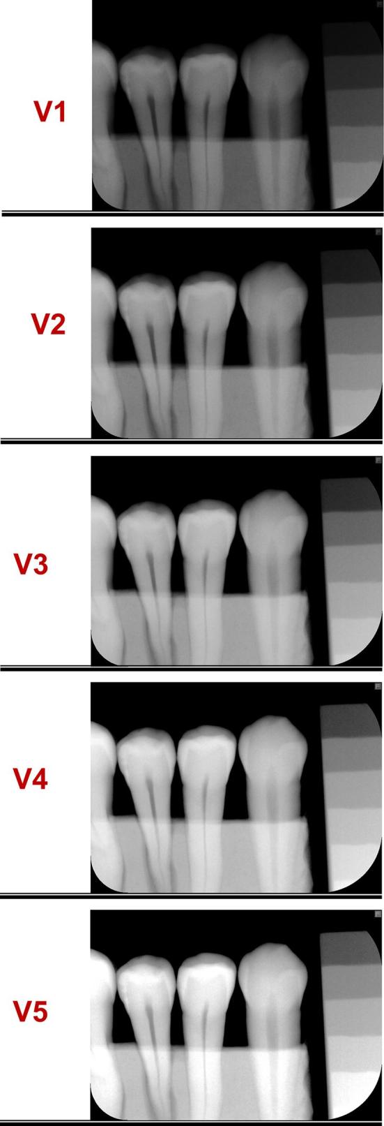
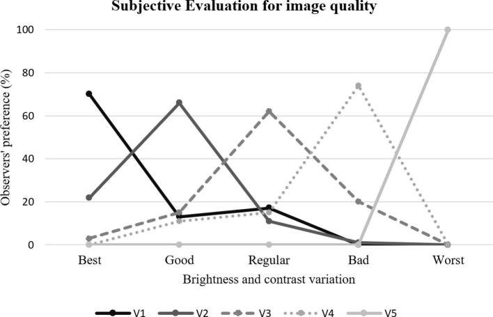
Methods: 80 proximal surfaces of posterior teeth were radiographed using an intraoral digital system (Digora Toto, Soredex, Finland). Initial images and four different combinations of brightness and contrast for each radiography were analysed. Five observers scored the images for the presence and extension of caries lesions. Micro-CT images were used as gold standard. In a second stage, the observers were asked which of the radiographs they preferred for the assessment of caries lesions.
Results: No differences were found between the original and adjusted radiographic images regarding the area under the receiver operating characteristic curve, sensitivity, and specificity (p > 0.05). There was a significant difference between the micro-CT and the intraoral radiographs (p < 0.0001). Images with high brightness and low contrast presented higher number of true negative cases, but also a decrease in caries detection. On the other hand, there were more cases of overestimation of the presence and extension of caries lesions in images with low brightness and high contrast. The subjective evaluation of image quality showed that radiographs with lower brightness and higher contrast tended to be preferred by observers.
Conclusions: Brightness and contrast adjustments in digital intraoral radiographs within the range tested in this study do not significantly influence the diagnosis of proximal caries lesions, although observers tend to prefer lower brightness and higher contrast images.
Figures
References
-
- World Health Organization (WHO). Sugars and dental caries. Geneva: WHO. 2017. Available from: http://apps.who.int/iris/bitstream/10665/259413/1/WHO-NMH-NHD-17.12-eng.pdf [accessed 26 January 2018].
-
- Kajan ZD, Tayefeh Davalloo R, Tavangar M, Valizade F. The effects of noise reduction, sharpening, enhancement, and image magnification on diagnostic accuracy of a photostimulable phosphor system in the detection of non-cavitated approximal dental caries. Imaging Sci Dent 2015; 45: 81–7. doi: 10.5624/isd.2015.45.2.81 - DOI - PMC - PubMed
MeSH terms
LinkOut - more resources
Full Text Sources
Other Literature Sources
Medical
