

Association of the Gutta-Induced Microenvironment With Corneal Endothelial Cell Behavior and Demise in Fuchs Endothelial Corneal Dystrophy
- PMID: 29852040
- PMCID: PMC6142944
- DOI: 10.1001/jamaophthalmol.2018.2031
Association of the Gutta-Induced Microenvironment With Corneal Endothelial Cell Behavior and Demise in Fuchs Endothelial Corneal Dystrophy
Abstract
Importance: The number and size of guttae increase over time in Fuchs endothelial corneal dystrophy (FECD); however, the association between these physical parameters and disease pathogenesis is unclear.
Objective: To determine the role of guttae in corneal endothelial cell function.
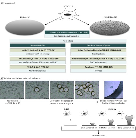
Design, settings, and participants: In an in vitro model, cells from a human corneal endothelial cell line, HCENC-21T, were seeded on decellularized normal (n = 30) and FECD (n = 70) endothelial basement (Descemet) membranes (DMs). Normal human corneas were sent to our laboratory from 3 sources. The study took place at the Schepens Eye Research Institute, Massachusetts Eye and Ear, Boston, and was performed from September 2015 to July 2017. Normal DMs were obtained from 3 different tissue banks and FECD-DMs were obtained from patients undergoing endothelial keratoplasty in 2 departments.
Main outcomes and measures: Endothelial cell shape, growth, and migration were assessed by live-cell imaging, and gene expression analysis as a function of guttae diameter was assessed by laser capture microscopy.
Results: Mean (SD) age of normal-DMs donors was 65.6 (4.4) years (16 women [53%]), and mean (SD) age of FECD-DMs donors was 68.9 (10.6) years (43 women [61%]). Cells covered a greater area (mean [SD], 97.7% [8.5%]) with a greater mean (SD) number of cells (2083 [153] cells/mm2) on the normal DMs compared with the FECD DMs (72.8% [11%]; P = .02 and 1541 [221] cells/mm2 221/mm2; P = .01, respectively). Differences in endothelial cell growth over guttae were observed on FECD DMs depending on the guttae diameter. Guttae with a mean (SD) diameter of 10.5 (2.9) μm did not impede cell growth, whereas those with a diameter of 21.1 (4.9) μm were covered only by the cell cytoplasm. Guttae with the largest mean (SD) diameter, 31.8 (3.8) μm, were not covered by cells, which instead surrounded them in a rosette pattern. Moreover, cells adjacent to large guttae upregulated αSMA, N-cadherin, Snail1, and NOX4 genes compared with ones grown on normal DMs or small guttae. Furthermore, large guttae induced TUNEL-positive apoptosis in a rosette pattern, similar to ex vivo FECD specimens.
Conclusions and relevance: These findings highlight the important role of guttae in endothelial cell growth, migration, and survival. These data suggest that cell therapy procedures in FECD might be guided by the diameter of the host guttae if subsequent clinical studies confirm these laboratory findings.
Conflict of interest statement
Figures
Comment in
-
The Ongoing Puzzle of the Biological Behavior of Cornea Guttae.JAMA Ophthalmol. 2018 Aug 1;136(8):893-894. doi: 10.1001/jamaophthalmol.2018.1475. JAMA Ophthalmol. 2018. PMID: 29852028 No abstract available.
References
-
- Joyce NC, Meklir B, Joyce SJ, Zieske JD. Cell cycle protein expression and proliferative status in human corneal cells. Invest Ophthalmol Vis Sci. 1996;37(4):645-655. - PubMed
-
- Joyce NC. Proliferative capacity of the corneal endothelium. Prog Retin Eye Res. 2003;22(3):359-389. - PubMed
-
- Bourne WM, Nelson LR, Hodge DO. Central corneal endothelial cell changes over a ten-year period. Invest Ophthalmol Vis Sci. 1997;38(3):779-782. - PubMed
Publication types
MeSH terms
Substances
Grants and funding
LinkOut - more resources
Full Text Sources
Other Literature Sources
Research Materials
Miscellaneous
