Review
. 2018 Jul 8:41:453-473.
doi: 10.1146/annurev-neuro-080317-061522. Epub 2018 May 31.

Endogenous and Exogenous Opioids in Pain

Affiliations
Review

Endogenous and Exogenous Opioids in Pain

Gregory Corder et al. Annu Rev Neurosci. .

Abstract

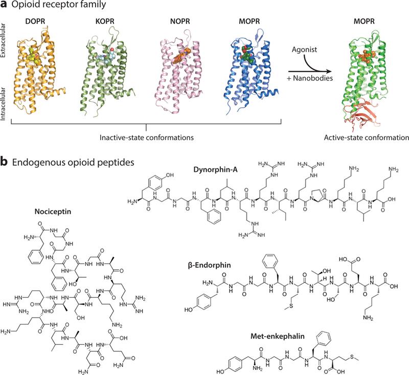
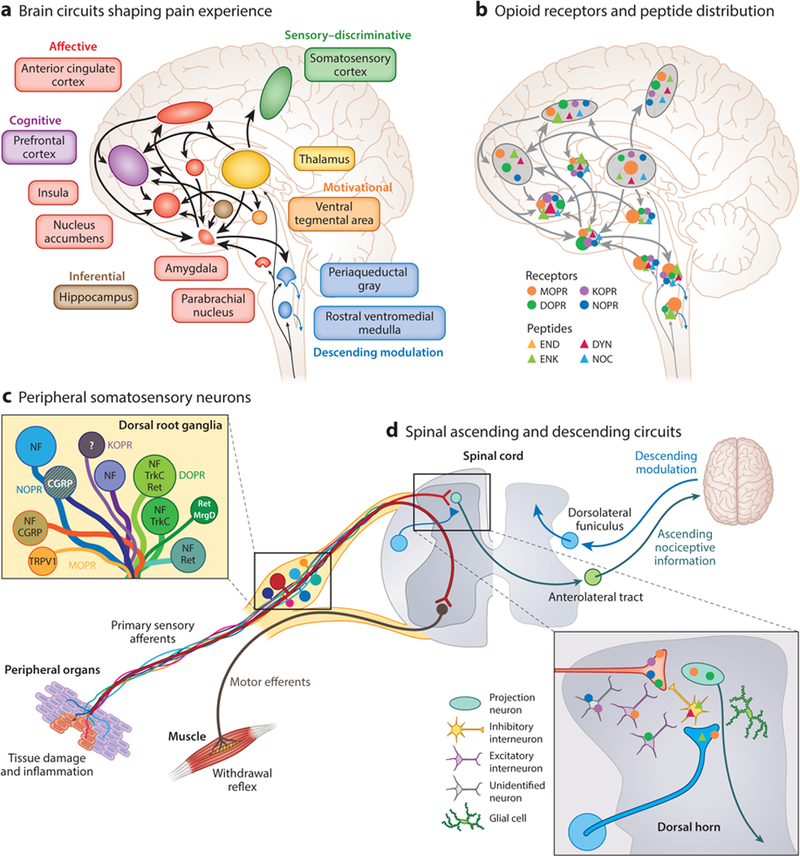
Opioids are the most commonly used and effective analgesic treatments for severe pain, but they have recently come under scrutiny owing to epidemic levels of abuse and overdose. These compounds act on the endogenous opioid system, which comprises four G protein-coupled receptors (mu, delta, kappa, and nociceptin) and four major peptide families (β-endorphin, enkephalins, dynorphins, and nociceptin/orphanin FQ). In this review, we first describe the functional organization and pharmacology of the endogenous opioid system. We then summarize current knowledge on the signaling mechanisms by which opioids regulate neuronal function and neurotransmission. Finally, we discuss the loci of opioid analgesic action along peripheral and central pain pathways, emphasizing the pain-relieving properties of opioids against the affective dimension of the pain experience.

Keywords: analgesia; neuroanatomy; opioid; pain; perception; signaling.


Figures

Figure 1
Figure 1
The endogenous opioid system. (a) Crystal structures of the inactive state of all four opioid receptors (DOPR, KOPR, NOPR, and MOPR). When an opioid agonist enters the binding pocket of its cognate receptor, a conformational change in the transmembrane domains allows for intracellular effector molecules to bind and activate signaling cascades that modulate neural function. The addition of stabilizing nanobodies to the crystal preparation has elucidated the active state of MOPR. Images courtesy of Dr. Aashish Manglik (UCSF) and used with his permission. (b) Chemical structures of the four main classes of opioid peptides: met-enkephalin, dynorphin-A, nociceptin, and β -endorphin. Abbreviations: DOPR, delta opioid receptor; KOPR, kappa opioid receptor; MOPR, mu opioid receptor; NOPR, nociceptin opioid receptor.
Figure 2
Figure 2
Opioid modulation of signaling and synaptic transmission. (a) Presynaptic and postsynaptic effects of opioids on nociception. (Left) Noxious stimuli trigger action potential firing along DRG nociceptors. Upon reaching the synaptic terminal, VGCCs (yellow) open, facilitating neurotransmitter release. These neurotransmitters (e.g., glutamate) then open postsynaptic AMPA and NMDA receptors, which continue the nociceptive signals along pain circuits. (Right) Activation of opioid receptors promotes dissociation of inhibitory Gα and Gβγ protein subunits. Gα subunits suppress adenylate cyclase, and Gβγ subunits presynaptically inhibit VGCC opening and postsynaptically activate GIRK channels, resulting in reduced neurotransmitter release and membrane hyperpolarization, respectively. (b) Biased signaling pathways. Agonist binding to opioid receptors causes conformational changes that promote distinct recruitment of G protein and arrestin effector signaling cascades. While G proteins mediate the inhibitory action of opioid signaling on neurotransmission, arrestin signaling is required both for internalization of opioid receptors and for kinase activities. The balance between G protein and arrestin signaling is thought, in part, to determine the analgesic versus detrimental effects of opioids. (c) Within pain circuits opioid receptors are activated by opioid analgesics such as enkephalin (endogenous) or morphine (exogenous). Endogenous opioids, such as enkephalins, can be released from infiltrating immune cells at the site of injuries and from neurons in the central nervous system. Abbreviations: AMPA, α-amino-3-hydroxy-5-methyl-4-isoxazolepropionic acid; DRG, dorsal root ganglion; EPSC, excitatory postsynaptic current; ERK, extracellular signal regulated kinase; GIRK, G protein gated inwardly rectifying potassium; JNK, c-Jun N-terminal kinase; NMDA, N-methyl-D-aspartate; RVM, rostral ventromedial medulla; VGCC, voltage-gated calcium channel.
Figure 3
Figure 3
Neuroanatomical substrates of pain perception and remodeling by opioids. (a) A large interconnected neural network of supraspinal brain circuits transforms nociceptive information ascending from the spinal cord into an aversive, painful experience. (b) The opioid system is well positioned within this brain network to modify the perception of pain. The different opioid receptors and peptides are distinctively, though broadly, expressed in different sites, the function of which is under intense investigation. Relative opioid receptor (circles) and peptide (triangles) expression levels are denoted by the size of the shapes. (c,d) Opioid receptor types and peptides are also distributed in distinct subpopulations of (c) DRG neurons, identified with the indicated markers such as TRPV1, and (d) second-order spinal cord dorsal horn neurons. NF marks large-diameter DRG neurons with myelinated axons. Striped neurons coexpress different opioid receptor types. Abbreviations: CGRP, calcitonin gene-related peptide; DRG, dorsal root ganglion; DOPR, delta opioid receptor; DYN, dynorphin; END, p-endorphin; ENK, enkephalin; KOPR, kappa opioid receptor; MOPR, mu opioid receptor; MrgD, Mas-related G protein-coupled receptor member D; NF, neurofilament; NOC, nociceptin/orphanin FQ; NOPR, nociceptin opioid receptor; Ret, Ret proto-oncogene; TrkC, tropomyosin receptor kinase C; TRPV1, transient receptor potential cation channel subfamily V member 1.

References

    1. Aicher SA, Punnoose A, Goldberg A. 2000. μ.-Opioid receptors often colocalize with the substance P receptor (NK1) in the trigeminal dorsal horn. J. Neurosci. 20:4345–54 - PMC - PubMed
    1. Al-Hasani R, Bruchas MR. 2011. Molecular mechanisms of opioid receptor-dependent signaling and behavior. Anesthesiology 115:1363–81 - PMC - PubMed
    1. Al-Hasani R, McCall JG, Shin G, Gomez AM, Schmitz GP, et al. 2015. Distinct subpopulations of nucleus accumbens dynorphin neurons drive aversion and reward. Neuron 87:1063–77 - PMC - PubMed
    1. al-Rodhan NR, Yaksh TL, Kelly PJ. 1992. Comparison of the neurochemistry of the endogenous opioid systems in two brainstem pain-processing centers. Stereotact. Funct. Neurosurg. 59:15–19 - PubMed
    1. Altier C, Khosravani H, Evans RM, Hameed S, Peloquin JB, et al. 2006. ORL1 receptor-mediated internalization of N-type calcium channels. Nat. Neurosci. 9:31–40 - PubMed

Publication types

Substances