

Mathematical models of dorsal closure
- PMID: 29852207
- PMCID: PMC6109426
- DOI: 10.1016/j.pbiomolbio.2018.05.009
Mathematical models of dorsal closure
Abstract
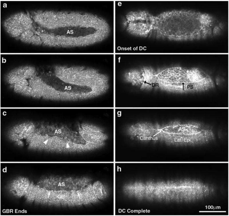
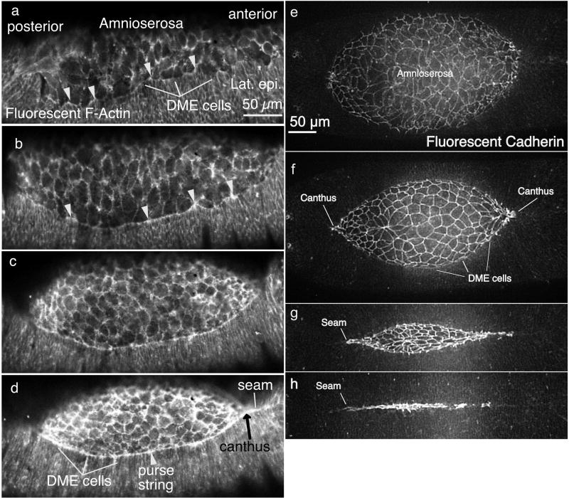
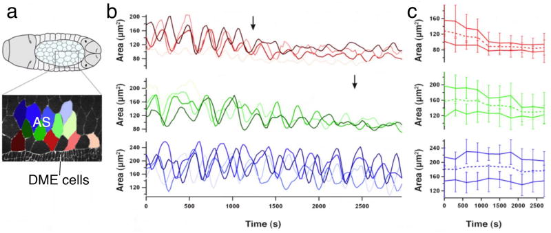
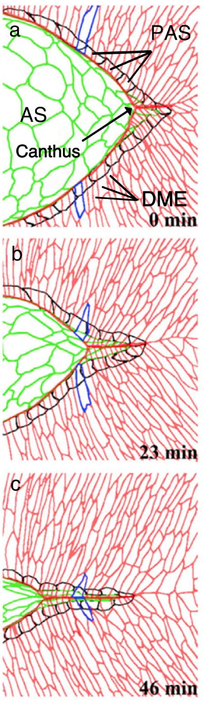
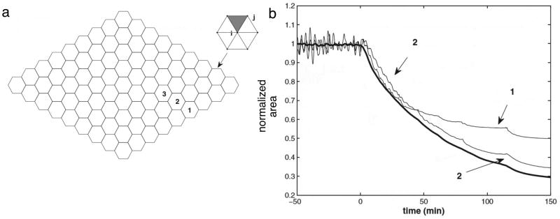
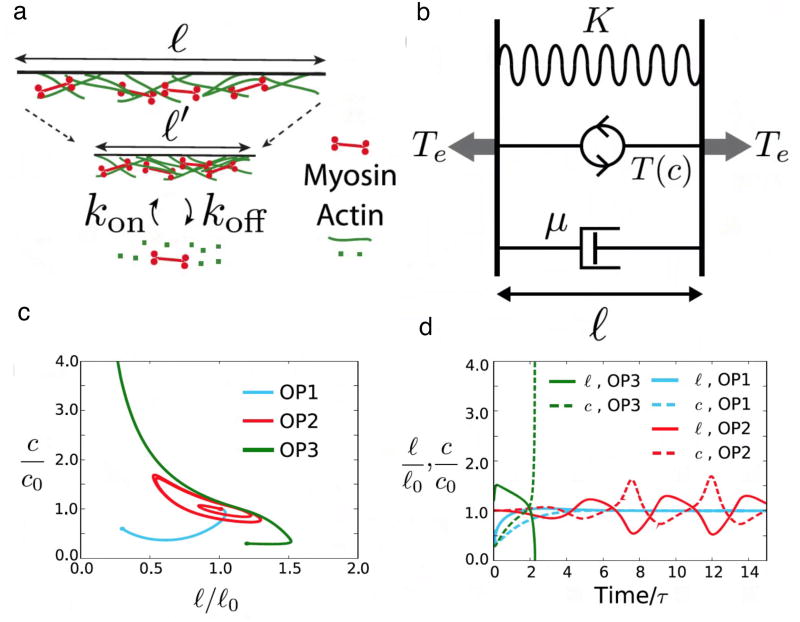
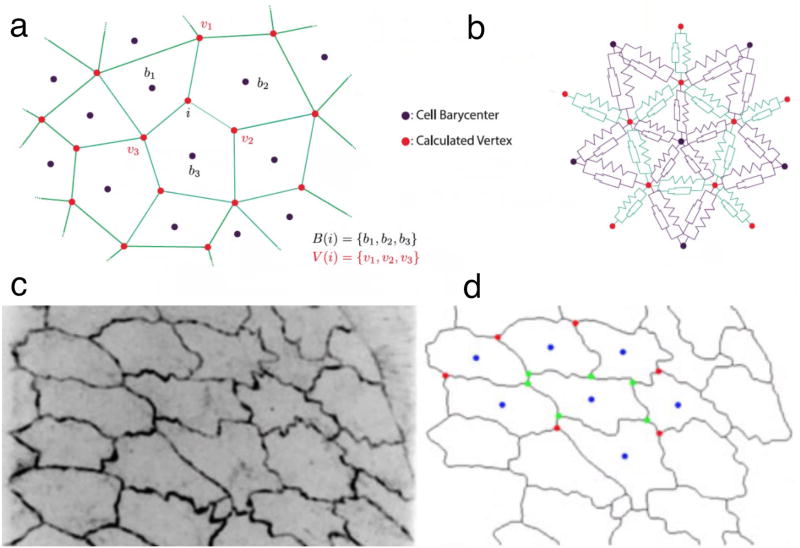
Dorsal closure is a model cell sheet movement that occurs midway through Drosophila embryogenesis. A dorsal hole, filled with amnioserosa, closes through the dorsalward elongation of lateral epidermal cell sheets. Closure requires contributions from 5 distinct tissues and well over 140 genes (see Mortensen et al., 2018, reviewed in Kiehart et al., 2017 and Hayes and Solon, 2017). In spite of this biological complexity, the movements (kinematics) of closure are geometrically simple at tissue, and in certain cases, at cellular scales. This simplicity has made closure the target of a number of mathematical models that seek to explain and quantify the processes that underlie closure's kinematics. The first (purely kinematic) modeling approach recapitulated well the time-evolving geometry of closure even though the underlying physical principles were not known. Almost all subsequent models delve into the forces of closure (i.e. the dynamics of closure). Models assign elastic, contractile and viscous forces which impact tissue and/or cell mechanics. They write rate equations which relate the forces to one another and to other variables, including those which represent geometric, kinematic, and or signaling characteristics. The time evolution of the variables is obtained by computing the solution of the model's system of equations, with optimized model parameters. The basis of the equations range from the phenomenological to biophysical first principles. We review various models and present their contribution to our understanding of the molecular mechanisms and biophysics of closure. Models of closure will contribute to our understanding of similar movements that characterize vertebrate morphogenesis.
Keywords: Actin; Cell junctions; Cell sheet morphogenesis; Computational; Myosin; Oscillation.
Copyright © 2018 Elsevier Ltd. All rights reserved.
Figures
Similar articles
-
Multiple forces contribute to cell sheet morphogenesis for dorsal closure in Drosophila.J Cell Biol. 2000 Apr 17;149(2):471-90. doi: 10.1083/jcb.149.2.471. J Cell Biol. 2000. PMID: 10769037 Free PMC article.
-
Cell Sheet Morphogenesis: Dorsal Closure in Drosophila melanogaster as a Model System.Annu Rev Cell Dev Biol. 2017 Oct 6;33:169-202. doi: 10.1146/annurev-cellbio-111315-125357. Annu Rev Cell Dev Biol. 2017. PMID: 28992442 Free PMC article. Review.
-
Nonmuscle myosin II generates forces that transmit tension and drive contraction in multiple tissues during dorsal closure.Curr Biol. 2005 Dec 20;15(24):2208-21. doi: 10.1016/j.cub.2005.11.064. Curr Biol. 2005. PMID: 16360683
-
Identifying Genetic Players in Cell Sheet Morphogenesis Using a Drosophila Deficiency Screen for Genes on Chromosome 2R Involved in Dorsal Closure.G3 (Bethesda). 2018 Jul 2;8(7):2361-2387. doi: 10.1534/g3.118.200233. G3 (Bethesda). 2018. PMID: 29776969 Free PMC article.
-
Amnioserosa development and function in Drosophila embryogenesis: Critical mechanical roles for an extraembryonic tissue.Dev Dyn. 2016 May;245(5):558-68. doi: 10.1002/dvdy.24395. Epub 2016 Mar 15. Dev Dyn. 2016. PMID: 26878336 Review.
Cited by
-
Dynamics of PAR Proteins Explain the Oscillation and Ratcheting Mechanisms in Dorsal Closure.Biophys J. 2018 Dec 4;115(11):2230-2241. doi: 10.1016/j.bpj.2018.10.014. Epub 2018 Oct 24. Biophys J. 2018. PMID: 30446158 Free PMC article.
-
A minimal vertex model explains how the amnioserosa avoids fluidization during Drosophila dorsal closure.Proc Natl Acad Sci U S A. 2025 Jan 7;122(1):e2322732121. doi: 10.1073/pnas.2322732121. Epub 2024 Dec 30. Proc Natl Acad Sci U S A. 2025. PMID: 39793057 Free PMC article.
-
A Comparative Study of the Role of Formins in Drosophila Embryonic Dorsal Closure.Cells. 2022 May 4;11(9):1539. doi: 10.3390/cells11091539. Cells. 2022. PMID: 35563844 Free PMC article.
-
Superresolution microscopy reveals actomyosin dynamics in medioapical arrays.Mol Biol Cell. 2022 Sep 15;33(11):ar94. doi: 10.1091/mbc.E21-11-0537. Epub 2022 May 11. Mol Biol Cell. 2022. PMID: 35544300 Free PMC article.
-
Automated profiling of gene function during embryonic development.Cell. 2024 Jun 6;187(12):3141-3160.e23. doi: 10.1016/j.cell.2024.04.012. Epub 2024 May 16. Cell. 2024. PMID: 38759650 Free PMC article.
References
-
- Almeida L, Bagnerini P, Habbal A, Noselli S, Serman F. Tissue repair modeling. In: Matteo Novaga GO, editor. Singularities in Nonlinear Evolution Phenomena and Applications. Edizioni Della Normale; Pisa, Italy: 2009. pp. 27–46.
-
- Almeida L, Bagnerini P, Habbal A, Noselli S, Serman F. A mathematical model for dorsal closure. Journal of Theoretical Biology. 2011;268:105–119. - PubMed
-
- Anderson ARA, Chaplain MAJ, McDougall S. A hybrid discrete-continuum model of tumour induced angiogenesis. In: Jackson TL, editor. Modeling Tumor Vasculature: Molecular, Cellular, and Tissue Level Aspects and Implications. Springer New York; New York, NY: 2012. pp. 105–133.
-
- Belacortu Y, Paricio N. Drosophila as a model of wound healing and tissue regeneration in vertebrates. Developmental Dynamics. 2011;240:2379–2404. - PubMed
Publication types
MeSH terms
Grants and funding
LinkOut - more resources
Full Text Sources
Other Literature Sources
Molecular Biology Databases
