

Identification of galectin-3 as an autoantigen in patients with IgG4-related disease
- PMID: 29852256
- PMCID: PMC6265117
- DOI: 10.1016/j.jaci.2018.05.011
Identification of galectin-3 as an autoantigen in patients with IgG4-related disease
Abstract
Background: The antigenic trigger that drives expansion of circulating plasmablasts and CD4+ cytotoxic T cells in patients with IgG4-related disease (IgG4-RD) is presently unknown.
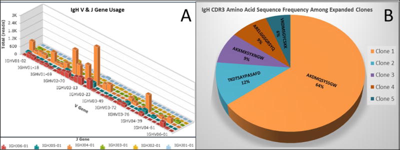
Objective: We sought to sequence immunoglobulin genes from single-cell clones of dominantly expanded plasmablasts and generate recombinant human mAbs to identify relevant antigens in patients with IgG4-RD by using mass spectrometry.
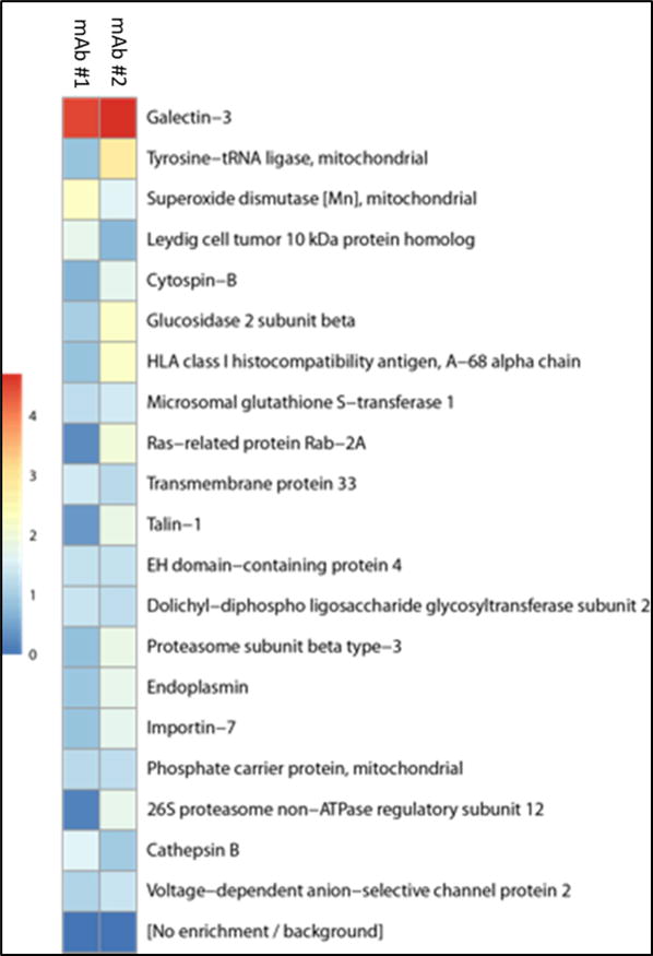
Methods: Paired heavy and light chain cDNAs from dominant plasmablast clones were expressed as mAbs and used to purify antigens by using immunoaffinity chromatography. Affinity-purified antigens were identified by using mass spectrometry and validated by means of ELISA. Plasma levels of the antigen of interest were also determined by using ELISA.
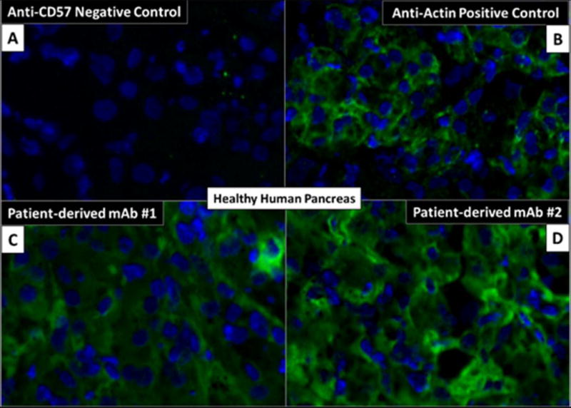
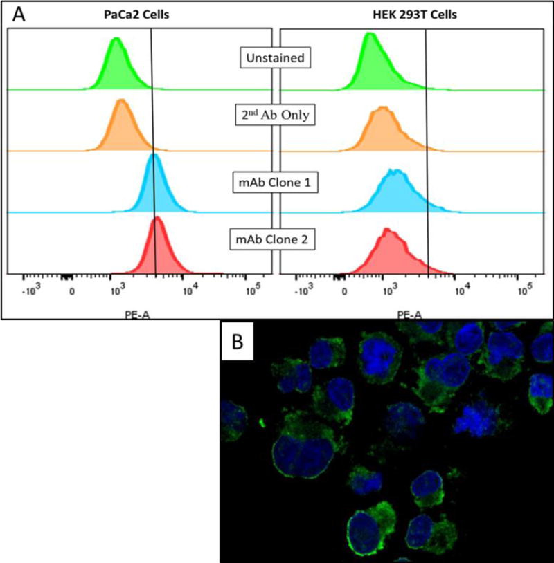
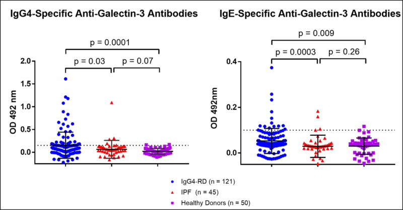
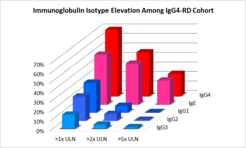
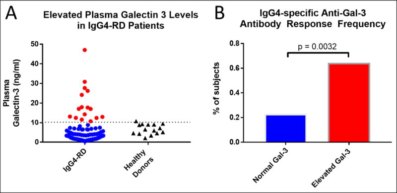
Results: mAbs expressed from the 2 dominant plasmablast clones of a patient with multiorgan IgG4-RD stained human pancreatic tissue sections. Galectin-3 was identified as the antigen specifically recognized by both mAbs. Anti-galectin-3 autoantibody responses were predominantly of the IgG4 isotype (28% of the IgG4-RD cohort, P = .0001) and IgE isotype (11% of the IgG4-RD cohort, P = .009). No significant responses were seen from the IgG1, IgG2, or IgG3 isotypes. IgG4 anti-galectin-3 autoantibodies correlated with increased plasma galectin-3 levels (P = .001), lymphadenopathy (P = .04), total IgG level increase (P = .05), and IgG4 level increase (P = .03).
Conclusion: Affinity chromatography using patient-derived mAbs identifies relevant autoantigens in patients with IgG4-RD. IgG4 galectin-3 autoantibodies are present in a subset of patients with IgG4-RD and correlate with galectin-3 plasma levels. The marked increases in levels of circulating IgG4 and IgE observed clinically are, at least in part, caused by the development of IgG4- and IgE-specific autoantibody responses.
Keywords: IgG(4)-related disease; autoantibody; autoantigen; galectin-3; plasmablast.
Copyright © 2018 American Academy of Allergy, Asthma & Immunology. Published by Elsevier Inc. All rights reserved.
Conflict of interest statement
The authors declare that they have no relevant conflict of interest.
Figures
Similar articles
-
Disease Severity Linked to Increase in Autoantibody Diversity in IgG4-Related Disease.Arthritis Rheumatol. 2020 Apr;72(4):687-693. doi: 10.1002/art.41140. Epub 2020 Feb 10. Arthritis Rheumatol. 2020. PMID: 31612628 Free PMC article.
-
The front line of research into immunoglobin G4-related disease - Do autoantibodies cause immunoglobin G4-related disease?Mod Rheumatol. 2019 Mar;29(2):214-218. doi: 10.1080/14397595.2018.1558519. Epub 2019 Jan 17. Mod Rheumatol. 2019. PMID: 30557067 Review.
-
De novo oligoclonal expansions of circulating plasmablasts in active and relapsing IgG4-related disease.J Allergy Clin Immunol. 2014 Sep;134(3):679-87. doi: 10.1016/j.jaci.2014.03.034. Epub 2014 May 6. J Allergy Clin Immunol. 2014. PMID: 24815737 Free PMC article.
-
Galectin-3 and prohibitin 1 are autoantigens in IgG4-related cholangitis without clear-cut protective effects against toxic bile acids.Front Immunol. 2024 Jan 25;14:1251134. doi: 10.3389/fimmu.2023.1251134. eCollection 2023. Front Immunol. 2024. PMID: 38332916 Free PMC article.
-
Pathogenesis of IgG4-related disease: a critical review.Odontology. 2019 Apr;107(2):127-132. doi: 10.1007/s10266-018-0377-y. Epub 2018 Jul 17. Odontology. 2019. PMID: 30019169 Review.
Cited by
-
Proteomic characteristics of saliva in patients with different subgroups of IgG4-RD.Front Immunol. 2022 Nov 22;13:1026921. doi: 10.3389/fimmu.2022.1026921. eCollection 2022. Front Immunol. 2022. PMID: 36483554 Free PMC article.
-
Immunoglobulin G4-related Disease.Clin Chest Med. 2019 Sep;40(3):583-597. doi: 10.1016/j.ccm.2019.05.005. Clin Chest Med. 2019. PMID: 31376893 Free PMC article. Review.
-
Current status of type 1 (IgG4-related) autoimmune pancreatitis.J Gastroenterol. 2022 Oct;57(10):695-708. doi: 10.1007/s00535-022-01891-7. Epub 2022 Aug 2. J Gastroenterol. 2022. PMID: 35916965 Free PMC article. Review.
-
A Case of Immunoglobulin G4-Related Disease Presenting as a Parapharyngeal Mass.Cureus. 2023 Jul 12;15(7):e41764. doi: 10.7759/cureus.41764. eCollection 2023 Jul. Cureus. 2023. PMID: 37575773 Free PMC article.
-
Review of a novel disease entity, immunoglobulin G4-related disease.J Korean Assoc Oral Maxillofac Surg. 2020 Feb;46(1):3-11. doi: 10.5125/jkaoms.2020.46.1.3. Epub 2020 Feb 26. J Korean Assoc Oral Maxillofac Surg. 2020. PMID: 32158675 Free PMC article. Review.
References
Publication types
MeSH terms
Substances
Grants and funding
LinkOut - more resources
Full Text Sources
Other Literature Sources
Research Materials
Miscellaneous
