. 2018 Nov 13;138(20):2247-2262.
doi: 10.1161/CIRCULATIONAHA.117.032821.

Beclin-1-Dependent Autophagy Protects the Heart During Sepsis

Affiliations

Beclin-1-Dependent Autophagy Protects the Heart During Sepsis

Yuxiao Sun et al. Circulation. .

Abstract

Background: Cardiac dysfunction is a major component of sepsis-induced multiorgan failure in critical care units. Changes in cardiac autophagy and its role during sepsis pathogenesis have not been clearly defined. Targeted autophagy-based therapeutic approaches for sepsis are not yet developed.

Methods: Beclin-1-dependent autophagy in the heart during sepsis and the potential therapeutic benefit of targeting this pathway were investigated in a mouse model of lipopolysaccharide (LPS)-induced sepsis.

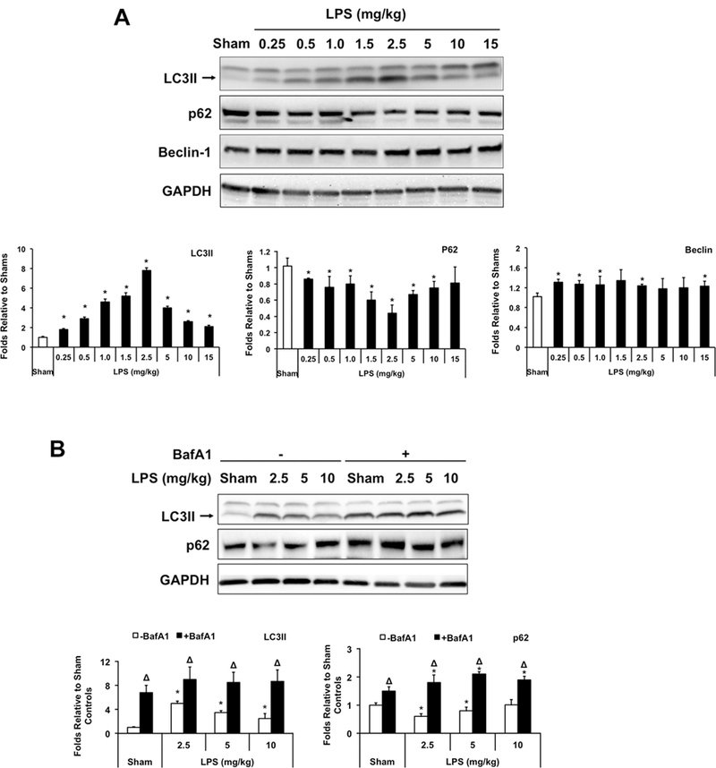
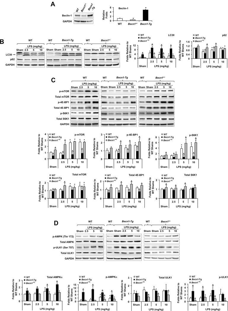
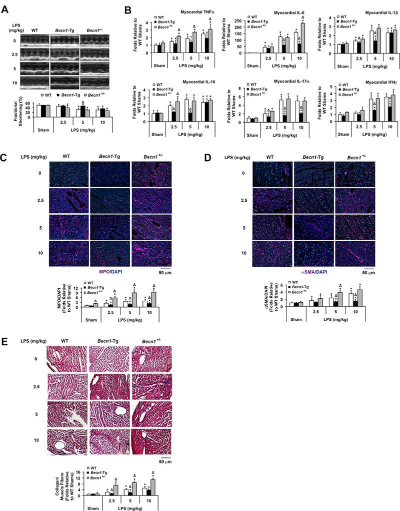
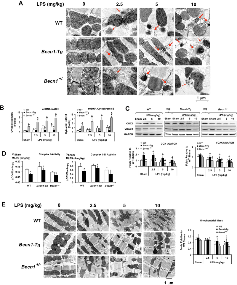
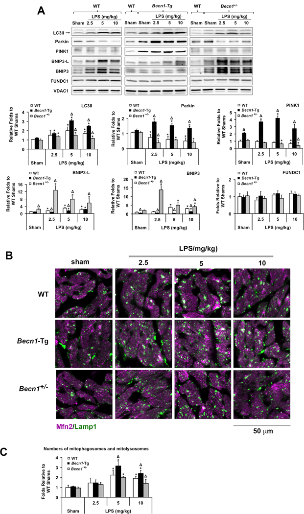
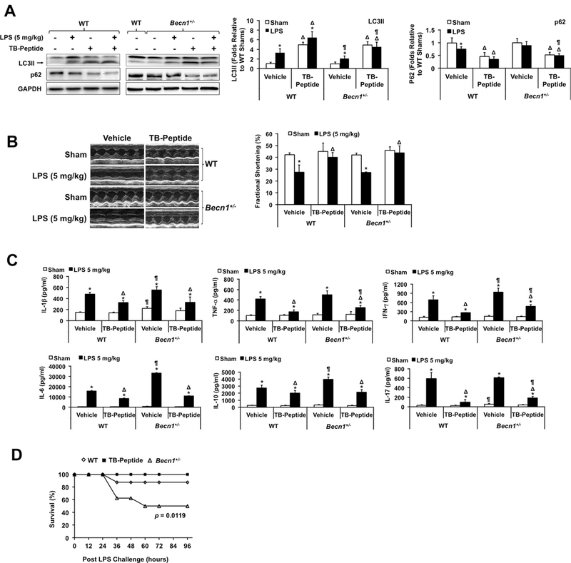
Results: LPS induced a dose-dependent increase in autophagy at low doses, followed by a decline that was in conjunction with mammalian target of rapamycin activation at high doses. Cardiac-specific overexpression of Beclin-1 promoted autophagy, suppressed mammalian target of rapamycin signaling, improved cardiac function, and alleviated inflammation and fibrosis after LPS challenge. Haplosufficiency for beclin 1 resulted in opposite effects. Beclin-1 also protected mitochondria, reduced the release of mitochondrial danger-associated molecular patterns, and promoted mitophagy via PTEN-induced putative kinase 1-Parkin but not adaptor proteins in response to LPS. Injection of a cell-permeable Tat-Beclin-1 peptide to activate autophagy improved cardiac function, attenuated inflammation, and rescued the phenotypes caused by beclin 1 deficiency in LPS-challenged mice.

Conclusions: These results suggest that Beclin-1 protects the heart during sepsis and that the targeted induction of Beclin-1 signaling may have important therapeutic potential.

Keywords: Beclin-1; autophagy; heart failure; mitochondrial degradation; sepsis.


Figures

Figure 1.
Figure 1.
Changes of cardiac autophagy in response to different doses of LPS. Wild-type C57BL/6 mice were given LPS via i.p. at indicated doses, heart tissues were harvested 18 hours later and total tissue lysates were prepared. A. Levels of LC3II, p62, Beclin and GAPDH were analyzed by Western blots using GAPDH as a loading control. B. LPS-induced autophagy flux was further confirmed by comparison of LC3II and p62 between animals with and without treatment of bafilomycin A1 (BafA1, 1.5 mg/kg). Results were quantified by densitometry and expressed as fold changes relative to shams. All values are means ± SEM. Significant differences are shown as * for sham vs. LPS-treated and Δ for vehicle vs. BafA1-treated groups (p < 0.05, n = 5, Mann-Whitney U test).
Figure 2.
Figure 2.
LPS-induced changes of cardiac autophagy and mTOR signaling in WT, Becn1-Tg, and Becn1+/− mice. Mice were given LPS via i.p. at indicated doses, heart tissues were harvested 18 hours later and total tissue lysates were prepared. Expression levels of Beclin-1 were confirmed (A), levels of LC3II and p62 (B), total and phosphorylated mTOR pathway molecules (C), and total and phosphorylated AMPKα and ULK1 (D) were examined by Western blots using GAPDH as a loading control. Results obtained by densitometry were expressed as fold changes relative to WT shams. All values are means ± SEM. Significant differences are shown as * for sham vs. LPS-treated and Δ for WT vs. Becn1-Tg or Becn1+/− groups (p < 0.05, n = 5, Mann-Whitney U test).
Figure 3.
Figure 3.
Evaluation of outcomes in LPS-challenged WT, Becn1-Tg, and Becn1+/− mice. Mice were given LPS via i.p. at indicated doses and analyzed 18 hours post challenge. A. Cardiac function was examined by echocardiography (n=5). B. Cytokines were measured in the heart tissue lysates by multiplex ELISA assays (n=5). C. Heart tissue sections were immune-stained with myeloperoxidase (MPO; purple) and co-stained with DAPI (blue) to show nucleus. D. Heart tissue sections were immune-stained with αSMA (purple) and co-stained with DAPI (blue) to show nucleus. E. Histological trichrome stain was applied to the heart tissue sections to visualize collagenous connective tissue fibers (blue). In C-E, images are representative of n ≥ 3 animals per group, and results were quantified using NIS Elements microscope imaging software. All values are means ± SEM. Significant differences are shown as * for sham vs. LPS-treated and Δ for WT vs. Becn1-Tg or Becn1+/− groups (p < 0.05, Mann-Whitney U test).
Figure 4.
Figure 4.
LPS-induced changes in mitochondrial structure and function in the heart of WT, Becn1-Tg, and Becn1+/− mice. Mice were given LPS via i.p. at indicated doses; heart tissues were harvested 18 hours later. A. Ultrastructure of myocardial mitochondria was observed by transmission electron microscope (EM). Red arrows indicated various stages of mitochondrial degradation. These images are representative of n≥3 animals per group. B. Total DNA was isolated from equal amount of cytosolic fractions and an exogenous internal positive control (IPC) DNA was spiked into all samples prior to DNA isolation as a positive control. Real-time PCR assays were performed using primers against mouse mtDNA NADH, cytochrome B or IPC. Results were expressed as a ratio of a target mtDNA to IPC. C. Levels of mtDNA encoded COX 1 and nuclear DNA encoded VDAC1 were examined in heart lysates by Western blots using GAPDH as a loading control. Results obtained by densitometry were expressed as fold changes relative to WT shams. D. Mitochondrial fractions were subjected to the measurements of complex I-V activities. E. Representative EM images of myocardial mitochondria at indicated magnitude. Mitochondrial mass was compared between groups. All values are means ± SEM. Significant differences are shown as * for sham vs. LPS-treated and Δ for WT vs. Becn1-Tg or Becn1+/− groups (p <0.05, n = 5, Mann-Whitney U test).
Figure 5.
Figure 5.
LPS-induced changes in mitophagy signaling molecules in the heart of WT, Becn1-Tg, and Becn1+/− mice. Mice were given LPS via i.p. at indicated doses, heart tissues were harvested 18 hours later. A. Levels of mitochondria-associated LC3II, Parkin, PINK1, BNIP3L, BNIP3, and FUNDC1 were examined by Western blots using VDAC1 as a loading control in mitochondrial fractions. Results obtained by densitometry were expressed as fold changes relative to WT shams. B. Heart tissue sections were co-stained with antibodies against Lamp1 (green) and Mfn2 (purple). Colors in white and pale green indicate the overlay of these two signals. Images are representative of n ≥ 3 animals per group. C. Number counts of mitophagosomes and mitolysosomes based on EM images. In A and C, all values are means ± SEM. Significant differences are shown as * for sham vs. LPS-treated and Δ for WT vs. Becn1-Tg or Becn1+/− groups (p < 0.05, n = 5, Mann-Whitney U test).
Figure 6.
Figure 6.
Effects of Tat-beclin-1 peptide (TB-peptide) in LPS-challenged mice. WT and Becn1+/− mice were given 5 mg/kg (A-C) or 10 mg/kg (D) LPS i.p. TB-peptide, 16 mg/kg, was administered i.p. 30 minutes post LPS challenge. Cardiac function was measured and tissues collected 18 hours post LPS challenge. A. Levels of LC3II, p62 and GAPDH were analyzed by Western blots using GAPDH as a loading control in heart tissue lysates. B. Cardiac function was examined by echocardiography. C. Cytokines in serum were measured by ELISA assays. In A-C, all values are means ± SEM. Significant differences are shown as * for with sham vs. LPS-treated, Δ for vehicle vs. TB-peptide-treated, and ¶ for WT vs. Becn1+/− groups (p < 0.05, n = 5, Mann-Whitney U test). D. Survival was monitored in LPS (10 mg/kg)-challenged WT ± TB-peptide and Becn1+/− mice, and * indicates a statistical significance when compared with WT (p < 0.05, n = 12, log-rank test).

Comment in

References

    1. Angus DC, Pereira CA and Silva E. Epidemiology of severe sepsis around the world. Endocr Metab Immune Disord Drug Targets. 2006;6:207–212. - PubMed
    1. Zanotti-Cavazzoni SL and Hollenberg SM. Cardiac dysfunction in severe sepsis and septic shock. Curr Opin Crit Care. 2009;15:392–397. - PubMed
    1. Kayar SR and Banchero N. Volume density and distribution of mitochondria in myocardial growth and hypertrophy. Respir Physiol. 1987;70:275–286. - PubMed
    1. Zang Q, Maass DL, Tsai SJ and Horton JW. Cardiac mitochondrial damage and inflammation responses in sepsis. Surg Infect (Larchmt). 2007;8:41–54. - PMC - PubMed
    1. Zang QS, Sadek H, Maass DL, Martinez B, Ma L, Kilgore JA, Williams NS, Frantz DE, Wigginton JG, Nwariaku FE, Wolf SE and Minei JP. Specific inhibition of mitochondrial oxidative stress suppresses inflammation and improves cardiac function in a rat pneumonia-related sepsis model. Am J Physiol Heart Circ Physiol. 2012;302:H1847–H1859. - PubMed

Publication types

MeSH terms