. 2018 Jul;38(7):1616-1631.
doi: 10.1161/ATVBAHA.118.311289. Epub 2018 May 31.

Deficiency of FAM3D (Family With Sequence Similarity 3, Member D), A Novel Chemokine, Attenuates Neutrophil Recruitment and Ameliorates Abdominal Aortic Aneurysm Development

Affiliations

Deficiency of FAM3D (Family With Sequence Similarity 3, Member D), A Novel Chemokine, Attenuates Neutrophil Recruitment and Ameliorates Abdominal Aortic Aneurysm Development

Li He et al. Arterioscler Thromb Vasc Biol. 2018 Jul.

Abstract

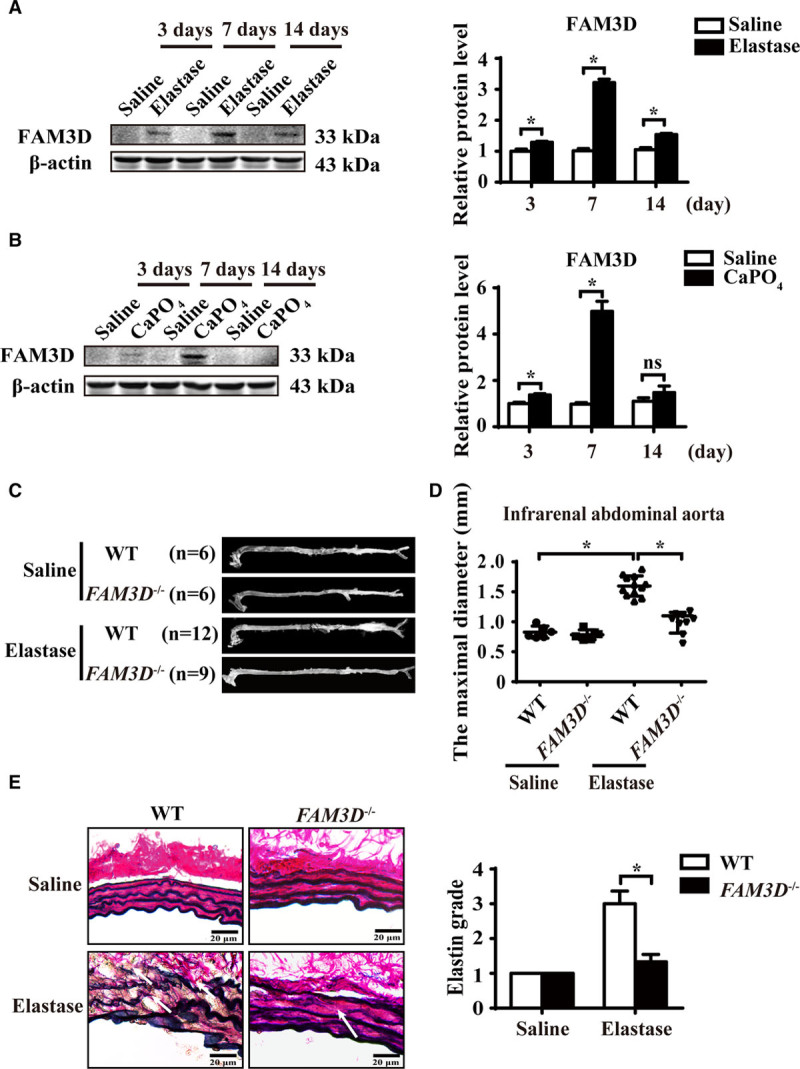
Objective: Chemokine-mediated neutrophil recruitment contributes to the pathogenesis of abdominal aortic aneurysm (AAA) and may serve as a promising therapeutic target. FAM3D (family with sequence similarity 3, member D) is a recently identified novel chemokine. Here, we aimed to explore the role of FAM3D in neutrophil recruitment and AAA development.

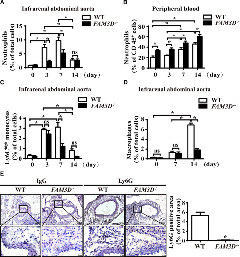
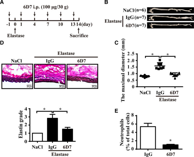
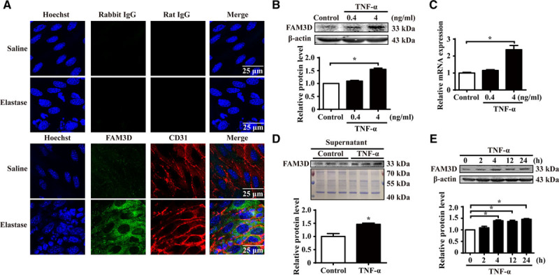
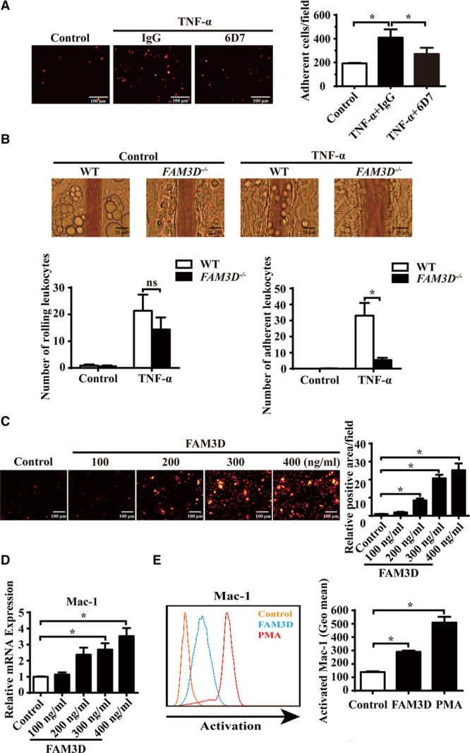
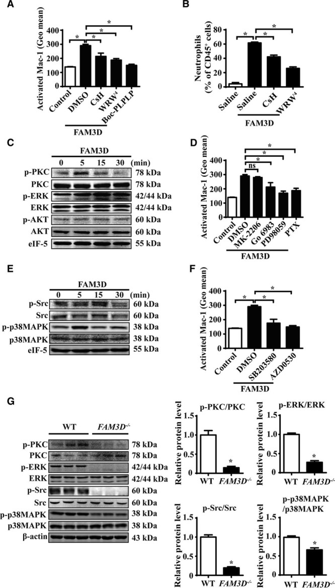
Approach and results: FAM3D was markedly upregulated in human AAA tissues, as well as both elastase- and CaPO4-induced mouse aneurysmal aortas. FAM3D deficiency significantly attenuated the development of AAA in both mouse models. Flow cytometry analysis indicated that FAM3D-/- mice exhibited decreased neutrophil infiltration in the aorta during the early stage of AAA formation compared with their wild-type littermates. Moreover, application of FAM3D-neutralizing antibody 6D7 through intraperitoneal injection markedly ameliorated elastase-induced AAA formation and neutrophil infiltration. Further, in vitro coculture experiments with FAM3D-neutralizing antibody 6D7 and in vivo intravital microscopic analysis indicated that endothelial cell-derived FAM3D induced neutrophil recruitment. Mechanistically, FAM3D upregulated and activated Mac-1 (macrophage-1 antigen) in neutrophils, whereas inhibition of FPR1 (formyl peptide receptor 1) or FPR2 significantly blocked FAM3D-induced Mac-1 activation, indicating that the effect of FAM3D was dependent on both FPRs. Moreover, specific inhibitors of FPR signaling related to Gi protein or β-arrestin inhibited FAM3D-activated Mac-1 in vitro, whereas FAM3D deficiency decreased the activation of both FPR-Gi protein and β-arrestin signaling in neutrophils in vivo.

Conclusions: FAM3D, as a dual agonist of FPR1 and FPR2, induced Mac-1-mediated neutrophil recruitment and aggravated AAA development through FPR-related Gi protein and β-arrestin signaling.

Keywords: G protein coupled receptor; abdominal aortic aneurysm; chemokine; endothelial cell; neutrophil recruitment.


Figures

Figure 1.
Figure 1.
FAM3D (family with sequence similarity 3, member D) deficiency attenuates the development of abdominal aortic aneurysm (AAA) in mice. A and B, Representative Western blot analysis and quantification of FAM3D expression in infrarenal abdominal aortas of C57BL/6 mice treated with elastase (A) or CaPO4 (B) for 3, 7, or 14 days. Saline was applied as a control. n=6. Two-way analysis of variance (ANOVA) followed by Tukey’s test for multiple comparisons, *P<0.05. C and D, Representative photographs (C) and quantification of maximal diameters (D) of the infrarenal abdominal aortas in saline- or elastase-induced 11- to 12-week-old wild-type (WT) and FAM3D−/− male mice. Two-way ANOVA followed by Tukey’s test for multiple comparisons, *P<0.05. E, Representative Verhoeff-Van Gieson staining and statistical analysis of elastin degradation in the infrarenal abdominal aortas in saline- or elastase-induced 11- to 12-week-old WT and FAM3D−/− male mice (n=6 for each group). Scale bar, 20 μm. Kruskal-Wallis test, *P<0.05.
Figure 2.
Figure 2.
FAM3D (family with sequence similarity 3, member D) deficiency reduces neutrophil recruitment to the aortic wall during abdominal aortic aneurysm (AAA) formation. A, Quantification of neutrophils in the infrarenal abdominal aortas of elastase-treated wild-type (WT) and FAM3D−/− mice at 0 day (WT, n=6; FAM3D−/−, n=6), 3 days (WT, n=7; FAM3D−/−, n=7), 7 days (WT, n=7; FAM3D−/−, n=6), and 14 days (WT, n=9; FAM3D−/−, n=7). Two-way analysis of variance (ANOVA) followed by Tukey’s test for multiple comparisons, *P<0.05, ns, no significance. B, Quantification of neutrophils in the peripheral blood of elastase-induced WT and FAM3D−/− mice at 0 day (WT, n=6; FAM3D−/−, n=6), 3 days (WT, n=7; FAM3D−/−, n=7), 7 days (WT, n=7; FAM3D−/−, n=6), and 14 days (WT, n=9; FAM3D−/−, n=7). Two-way ANOVA followed by Tukey’s test for multiple comparisons, *P<0.05. C, Quantification of Ly6Chigh monocytes in the infrarenal abdominal aortas of elastase-treated WT and FAM3D−/− mice at 0 day (WT, n=6; FAM3D−/−, n=6), 3 days (WT, n=7; FAM3D−/−, n=7), 7 days (WT, n=7; FAM3D−/−, n=7), and 14 days (WT, n=7; FAM3D−/−, n=7). Two-way ANOVA followed by Tukey’s test for multiple comparisons, *P<0.05, ns, no significance. D, Quantification of macrophages in infrarenal abdominal aortas of elastase-treated WT and FAM3D−/− mice at 0 day (WT, n=6; FAM3D−/−, n=6), 7 days (WT, n=7; FAM3D−/−, n=6), and 14 days (WT, n=9; FAM3D-/-, n=7). Two-way ANOVA followed by Tukey’s test for multiple comparisons, *P<0.05, ns, no significance. E, Representative immunohistochemical staining and quantification of neutrophil infiltration in infrarenal abdominal aortas of WT and FAM3D−/− mice 7 days after elastase induction, using the neutrophil-specific antibody Ly6G. Rat IgG was applied as a negative control. WT, n=8; FAM3D−/−, n=7. Scale bar, 20 μm. Mann-Whitney test, *P<0.05.
Figure 3.
Figure 3.
FAM3D (family with sequence similarity 3, member D) neutralization antibody 6D7 attenuates the development of abdominal aortic aneurysm (AAA) in mice. A, Schematic illustration of the time points of intraperitoneal injection of 6D7 or mouse IgG (100 μg/30 g body weight). B and C, Representative photographs (B) and quantification of maximal diameters (C) of the infrarenal abdominal aortas of 11- to 12-week-old C57BL/6 male mice treated with elastase for 14 days along with mouse IgG or 6D7 injection (NaCl, n=6; elastase+IgG, n=7; elastase+6D7, n=7). One-way ANOVA followed by Tukey’s test for multiple comparisons, *P<0.05. D, Representative Verhoeff-Van Gieson staining and statistical analysis of elastin degradation in the infrarenal abdominal aortas of 11- to 12-week-old C57BL/6 male mice treated with elastase for 14 days along with mouse IgG or 6D7 injection (NaCl, n=6; elastase+IgG, n=7; elastase+6D7, n=7). Scale bar, 20 μm. Kruskal-Wallis test, *P<0.05. E, Quantification of neutrophils in the infrarenal abdominal aortas of wild-type (WT) mice treated with elastase for 7 days along with mouse IgG or 6D7 injection (IgG, n=7; 6D7, n=7). Unpaired Student t test, *P<0.05.
Figure 4.
Figure 4.
FAM3D (family with sequence similarity 3, member D) is expressed and upregulated in endothelial cells. A, Representative immunofluorescent en face staining of FAM3D expression in the endothelial layer of infrarenal abdominal aortas from 11- to 12-week-old male C57BL/6 mice 7 days after saline (n=6) or elastase (n=6) treatment. Rabbit IgG and rat IgG were used as the negative controls of FAM3D and CD31 antibodies, respectively. B, Representative Western blot analysis and quantification of FAM3D expression in human umbilical vein endothelial cells (HUVECs) treated with TNF-α (tumor necrosis factor α) for 24 hours. n=3. One-way analysis of variance (ANOVA) followed by Tukey’s test for multiple comparisons, *P<0.05. C, Real-time polymerase chain reaction (PCR) analysis of FAM3D expression in HUVECs treated with TNF-α for 24 hours. β-Actin was applied as an internal control. n=6. One-way ANOVA followed by Tukey’s test for multiple comparisons, *P<0.05. D, Representative Western blot analysis and quantification of FAM3D in the supernatant of HUVECs stimulated by TNF-α (4 ng/mL) for 24 hours. Coomassie blue staining of the gel loaded with cell lysates of HUVECs was applied as the internal control. n=3. Unpaired Student t test, *P<0.05. E, Representative Western blot analysis and quantification of FAM3D expression in HUVECs treated with TNF-α (4 ng/mL) for 0, 2, 4, 12, and 24 hours. n=3. One-way ANOVA followed by Tukey’s test for multiple comparisons, *P<0.05.
Figure 5.
Figure 5.
FAM3D (family with sequence similarity 3, member D) induces neutrophil adhesion and transmigration and activates Mac-1 (macrophage-1 antigen) in neutrophils. A, Representative photographs and quantification of the adhesion of DiI-labeled neutrophils to human umbilical vein endothelial cells (HUVECs) after coculture in the absence or presence of FAM3D neutralization antibody 6D7 (20 μmol/L) or an equal amount of mouse IgG for 2 hours. HUVEC monolayers were stimulated with 4 ng/mL TNF-α (tumor necrosis factor α) for 24 hours prior to coculture. n=12. One-way analysis of variance (ANOVA) followed by Tukey’s test for multiple comparisons, *P<0.05. B, Representative intravital microscopic photographs and quantification of leukocyte rolling and adhesion to mesentery microvessels of wild-type (WT; n=8) and FAM3D−/− (n=8) mice 4 hours after intraperitoneal injection with or without TNF-α (500 ng for each mice). Two-way ANOVA followed by Tukey’s test for multiple comparisons, *P<0.05, ns, no significance. C, Representative photographs and quantification of DiI-stained mouse neutrophils transmigrated through a confluent layer of HUVECs in response to 4 hours of treatment with mouse FAM3D. n=12. One-way ANOVA followed by Tukey’s test for multiple comparisons, *P<0.05. D, Real-time polymerase chain reaction (PCR) quantification of mRNA levels of Mac-1 (CD11b) in mouse neutrophils stimulated by FAM3D for 4 hours. β-Actin was applied as an internal control. n=6. One-way ANOVA followed by Tukey’s test for multiple comparisons, *P<0.05. E, Representative flow cytometric analysis and quantification of activated Mac-1 (CD11b) induced by FAM3D (10 nmol/L) or phorbol-12-myristate-13-acetate (PMA; 10 nmol/L) in human neutrophils. n=6. One-way ANOVA followed by Tukey’s test for multiple comparisons, *P<0.05, ns, no significance.
Figure 6.
Figure 6.
FAM3D (family with sequence similarity 3, member D) induces Mac-1 (macrophage-1 antigen) activation via FPR (formyl peptide receptor)-Gi protein/β-arrestin signaling. A, Flow cytometric quantification of activated Mac-1 (CD11b) in human neutrophils pretreated with 10 ng/mL of Boc-PLPLP, 5 μmol/L of cyclosporine H (CsH), or 5 μmol/L of WRW for 2 hours followed by FAM3D (400 ng/mL) treatment for another 15 minutes. n=6. One-way analysis of variance (ANOVA) followed by Tukey’s test for multiple comparisons, *P<0.05. B, C57BL/6 mice (11–12 weeks old) underwent the intraperitoneal injection of CsH (10 μg/30 g body weight) or WRW (10 μg/30 g body weight) at 1 hour prior to FAM3D induction. Then FAM3D (10 μg/30 g body weight) was injected into peritoneal cavity for neutrophil recruitment in 6 hours. The percentage of neutrophils in CD45+ peritoneal cells was analyzed as CD45+CD11b+Ly6G+ by flow cytometry. n=7. One-way ANOVA followed by Tukey’s test for multiple comparisons, *P<0.05. C, Representative Western blot analysis of PKC (protein kinase C), ERK (extracellular regulated MAP kinase), and AKT (PKB, protein kinase B) activation in human neutrophils treated with 400 ng/mL FAM3D for various periods. D, Human neutrophils were pretreated with 20 μmol/L of MK-2206, 100 nmol/L of Go6983, 10 μmol/L of PD98059 or 10 ng/mL of PTX for 2 hours, followed by FAM3D (400 ng/mL) treatment for another 15 minutes, and the activation of Mac-1 (CD11b) was analyzed by flow cytometric assay. n=6. One-way analysis of variance (ANOVA) followed by Tukey’s test for multiple comparisons, *P<0.05, ns, no significance. E, Representative Western blot analysis of Src and p38MAPK (mitogen-activated kinase-like protein) activation in human neutrophils treated with 400 ng/mL FAM3D for various periods. F, Human neutrophils were pretreated with 10 μmol/L of SB203580 or 5 μmol/L of AZD0530, respectively, for 2 hours followed by FAM3D (400 ng/mL) treatment for another 15 minutes; the activation of Mac-1 was analyzed by flow cytometric assay. n=6. One-way ANOVA followed by Tukey’s test for multiple comparisons, *P<0.05. G, Representative Western blot analysis and quantification of PKC, ERK, Src, and p38MAPK in peripheral blood neutrophils isolated from wild-type (WT; n=6) and FAM3D−/− (n=6) mice treated with elastase for 7 days. Unpaired Student t test, *P<0.05. HUVECs indicates human umbilical vein endothelial cells; PTX, pertussis toxin; and TNF-α, tumor necrosis factor α.
Figure 7.
Figure 7.
Schematic illustration of Mac-1 (macrophage-1 antigen) activation induced by FAM3D (family with sequence similarity 3, member D). Pathological stimuli including TNF-α, LPS, and OSS promote the expression and secretion of endothelial cell (EC)–generated FAM3D. FAM3D activates Mac-1 on neutrophils through FPR (formyl peptide receptor) 1 and FPR2, dependent on Gi-protein- and β-arrestin-related signals. The activated Mac-1 interacts with ICAM-1 on the endothelial surface, mediating neutrophil recruitment and abdominal aortic aneurysm (AAA) development. ICAM-1 indicates intercellular adhesion molecule 1; LPS, lipopolysaccharide; OSS, oscillatory shear stress; and TNF-α, tumor necrosis factor α.

References

    1. Johnston KW, Rutherford RB, Tilson MD, Shah DM, Hollier L, Stanley JC. Suggested standards for reporting on arterial aneurysms. Subcommittee on Reporting Standards for Arterial Aneurysms, Ad Hoc Committee on Reporting Standards, Society for Vascular Surgery and North American Chapter, International Society for Cardiovascular Surgery. J Vasc Surg. 1991;13:452–458. - PubMed
    1. Nordon IM, Hinchliffe RJ, Loftus IM, Thompson MM. Pathophysiology and epidemiology of abdominal aortic aneurysms. Nat Rev Cardiol. 2011;8:92–102. doi: 10.1038/nrcardio.2010.180. - PubMed
    1. Rizas KD, Ippagunta N, Tilson MD., III Immune cells and molecular mediators in the pathogenesis of the abdominal aortic aneurysm. Cardiol Rev. 2009;17:201–210. doi: 10.1097/CRD.0b013e3181b04698. - PubMed
    1. Buck DB, van Herwaarden JA, Schermerhorn ML, Moll FL. Endovascular treatment of abdominal aortic aneurysms. Nat Rev Cardiol. 2014;11:112–123. doi: 10.1038/nrcardio.2013.196. - PMC - PubMed
    1. Weber C, Zernecke A, Libby P. The multifaceted contributions of leukocyte subsets to atherosclerosis: lessons from mouse models. Nat Rev Immunol. 2008;8:802–815. doi: 10.1038/nri2415. - PubMed

Publication types

MeSH terms