. 2018 Jul 4;38(27):6063-6075.
doi: 10.1523/JNEUROSCI.3647-17.2018. Epub 2018 May 31.

Glial βII Spectrin Contributes to Paranode Formation and Maintenance

Affiliations

Glial βII Spectrin Contributes to Paranode Formation and Maintenance

Keiichiro Susuki et al. J Neurosci. .

Abstract

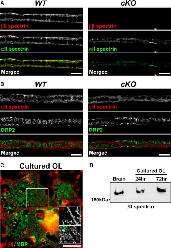
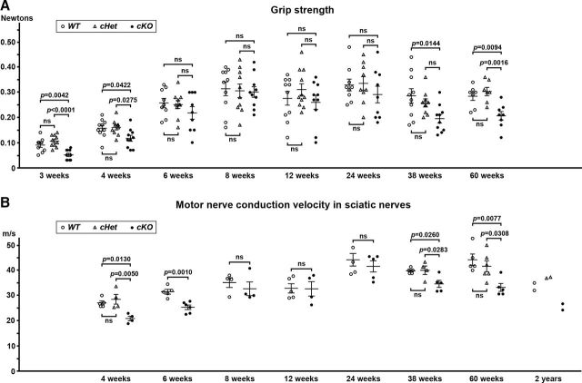
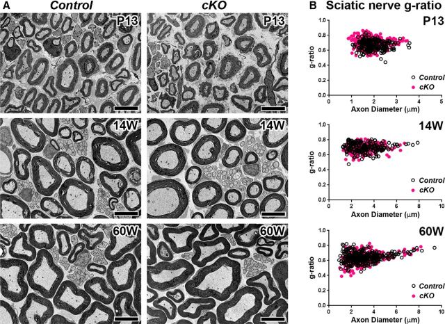
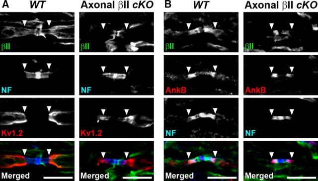
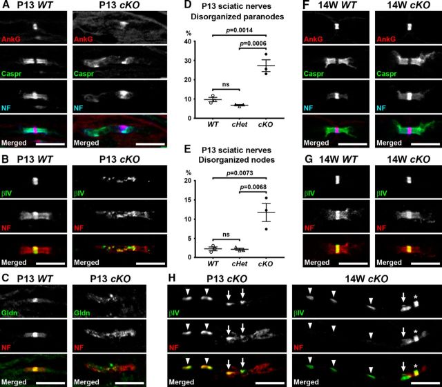
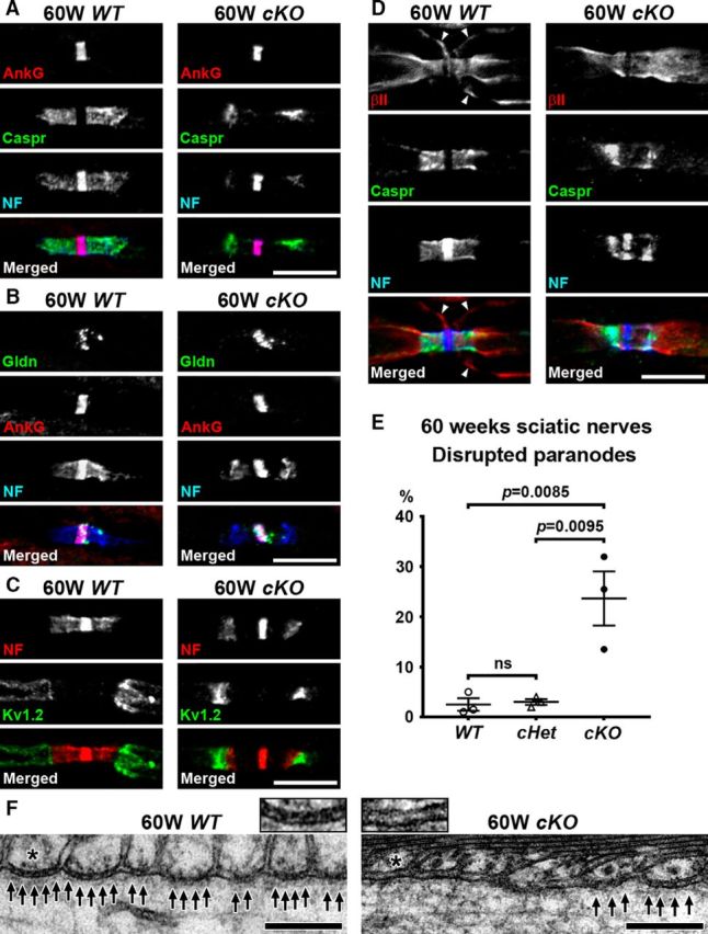
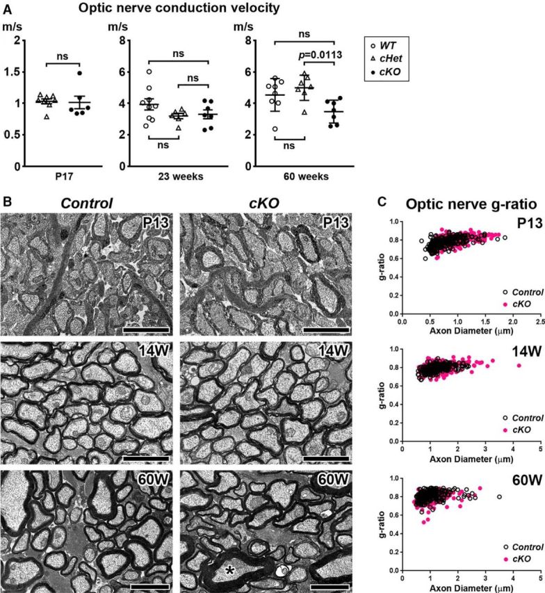
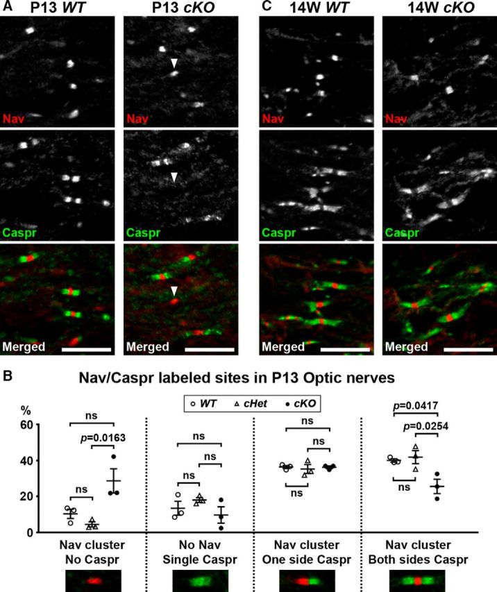
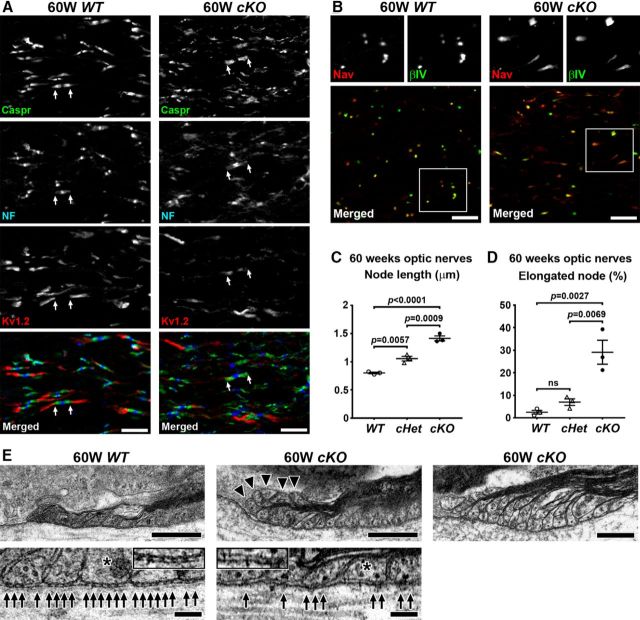
Action potential conduction along myelinated axons depends on high densities of voltage-gated Na+ channels at the nodes of Ranvier. Flanking each node, paranodal junctions (paranodes) are formed between axons and Schwann cells in the peripheral nervous system (PNS) or oligodendrocytes in the CNS. Paranodal junctions contribute to both node assembly and maintenance. Despite their importance, the molecular mechanisms responsible for paranode assembly and maintenance remain poorly understood. βII spectrin is expressed in diverse cells and is an essential part of the submembranous cytoskeleton. Here, we show that Schwann cell βII spectrin is highly enriched at paranodes. To elucidate the roles of glial βII spectrin, we generated mutant mice lacking βII spectrin in myelinating glial cells by crossing mice with a floxed allele of Sptbn1 with Cnp-Cre mice, and analyzed both male and female mice. Juvenile (4 weeks) and middle-aged (60 weeks) mutant mice showed reduced grip strength and sciatic nerve conduction slowing, whereas no phenotype was observed between 8 and 24 weeks of age. Consistent with these findings, immunofluorescence microscopy revealed disorganized paranodes in the PNS and CNS of both postnatal day 13 and middle-aged mutant mice, but not in young adult mutant mice. Electron microscopy confirmed partial loss of transverse bands at the paranodal axoglial junction in the middle-aged mutant mice in both the PNS and CNS. These findings demonstrate that a spectrin-based cytoskeleton in myelinating glia contributes to formation and maintenance of paranodal junctions.SIGNIFICANCE STATEMENT Myelinating glia form paranodal axoglial junctions that flank both sides of the nodes of Ranvier. These junctions contribute to node formation and maintenance and are essential for proper nervous system function. We found that a submembranous spectrin cytoskeleton is highly enriched at paranodes in Schwann cells. Ablation of βII spectrin in myelinating glial cells disrupted the paranodal cell adhesion complex in both peripheral and CNSs, resulting in muscle weakness and sciatic nerve conduction slowing in juvenile and middle-aged mice. Our data show that a spectrin-based submembranous cytoskeleton in myelinating glia plays important roles in paranode formation and maintenance.

Keywords: myelin; node of Ranvier; paranode; spectrin.


Figures

Figure 1.
Figure 1.
βII and αII spectrin expression and localization in myelinating Schwann cells and oligodendrocytes. A, B, Teased fiber preparation of sciatic nerves from Sptbn1f/f mice (WT, left) or Cnp-Cre;Sptbn1f/f mice (cKO, right) at 8 weeks of age. Nerves were labeled using antibodies against βII spectrin (red) and αII spectrin (A, green), or against αII spectrin (red) and DRP2 (B, green). Scale bars: A, B, 10 μm. C, Cultured rat oligodendrocyte (OL) immunostained for βII spectrin (red) and MBP (green). Scale bar, 20 μm. D, Immunoblotting of brain homogenates and protein extracts derived from purified OL cultures of 24 and 72 h for βII spectrin.
Figure 2.
Figure 2.
Phenotypes in mutant mice lacking βII spectrin in myelinating glial cells. A, Toe grip strength measurement. Grip strength in each mouse is plotted. N = 7–11 mice in each group. B, Motor nerve conduction velocity in sciatic nerves. Conduction velocity for each mouse is plotted. N = 4–6 mice in each group. Differences of conduction velocity between WT and cKO are shown in 6–24 weeks. Two mice at 2 years of age in each group were available for motor nerve conduction measurements. WT, Sptbn1f/f mice; cHet, Cnp-Cre;Sptbn1f /+ mice; and cKO, Cnp-Cre;Sptbn1f/f mice.
Figure 3.
Figure 3.
βII spectrin-deficient Schwann cell myelinated axons. A, Cross sections of sciatic nerves from Control (WT, Sptbn1f/f); and cKO, Cnp-Cre;Sptbn1f/f mice at P13, 14 weeks, and 60 weeks of age. Scale bars, 5 μm. B, Scatter plots of g-ratio (y-axis) in relation to axon diameter (x-axis) of individual fiber in sciatic nerves. g-ratios: Control (0.6693 ± 0.0617, mean ± SD; n = 219 axons) and cKO (0.7131 ± 0.0695, mean ± SD; n = 316 axons) at P13; Control (0.697 ± 0.0578, mean ± SD; n = 203 axons) and cKO (0.6871 ± 0.0588, mean ± SD; n = 187 axons) at 14 weeks of age; and Control (0.6447 ± 0.0653, mean ± SD; n = 315 axons) and cKO (0.6284 ± 0.0698, mean ± SD; n = 314 axons) at 60 weeks of age. Data were collected from two control (1 WT and 1 cHet) and three cKO mice at P13; two control (WT) and two cKO mice at 14 weeks, and three control (WT) and three cKO mice at 60 weeks of age. Approximately 100 axons were analyzed per mouse.
Figure 4.
Figure 4.
Schwann cell βII spectrin is at paranodes. Dorsal roots from WT (Sptbn1f/f) or mutant mice lacking βII spectrin in sensory axons (axonal βII cKO, Avil-Cre;Sptbn1f/f) were immunostained using antibodies to βII spectrin (green), NF (blue), and Kv1.2 (A, red), or antibodies to βII spectrin (green), AnkyrinB (AnkB; red), and NF (B, blue). Arrowheads indicate the boundary between paranodes and juxtaparanodes. Scale bars, 10 μm.
Figure 5.
Figure 5.
Altered paranode and node organization during early development in sciatic nerves lacking Schwann cell βII spectrin. AC, Sciatic nerve sections from P13 WT (Sptbn1f/f) and cKO (Cnp-Cre;Sptbn1f/f) mice immunostained for AnkG (red), Caspr (green), and NF (A, blue), βIV spectrin (βIV; green), and NF (B, red), or gliomedin (Gldn; green) and NF (C, red). Scale bars, 10 μm. D, E, The frequency of disorganized paranodes (dispersed or reduced NF155 staining; D) and nodes (dispersed βIV spectrin staining; E) in sciatic nerves from WT (Sptbn1f/f), cHet (Cnp-Cre;Sptbn1f /+), and cKO (Cnp-Cre;Sptbn1f/f) mice at P13. Data were collected from three mice in each group. In each mouse, two sciatic nerve sections were analyzed, and ~100 paranodes and nodes were observed in each section. F, G, Sciatic nerve sections from WT and cKO mice at 14 weeks of age immunostained for AnkG (red), Caspr (green), and NF (F, blue), or βIV spectrin (βIV; green) and NF (G, red). Scale bars, 10 μm. H, Sciatic nerve sections immunostained using antibodies against βIV spectrin (green) and NF (red). Ectopic βIV spectrin clusters are observed at paranodes (arrows) or at internodes (arrowheads) in both P13 (left column) and 14 weeks (right column) cKO sciatic nerves. Asterisk indicates a node. Scale bars, 10 μm.
Figure 6.
Figure 6.
Paranodal junctions are disrupted in middle-aged sciatic nerves lacking Schwann cell βII spectrin. AD, Nodes of Ranvier from WT (Sptbn1f/f) or cKO (Cnp-Cre;Sptbn1f/f) mice at 60 weeks of age are immunostained as indicated. Immunostaining of Caspr (green) and NF155 (blue) at paranodes is reduced in cKO nerves (AD). Nodal clusters of AnkG (A, B, red), gliomedin (Gldn; B, green), and NF186 (NF; AD, blue) are preserved. Juxtaparanodal Kv1.2 (green) is also preserved (C). The arrowheads in D indicate Schwann cell βII spectrin immunostaining near the outer surface of myelin sheath in WT nerve, but this Schwann cell βII spectrin staining is completely abolished in cKO nerve. Scale bars, 10 μm. E, Frequency of reduced paranodal Caspr and NF staining in sciatic nerves from WT (Sptbn1f/f), cHet (Cnp-Cre;Sptbn1f/+), and cKO (Cnp-Cre;Sptbn1f/f) mice at 60 weeks of age. N = 3 mice in each group. Paranodes were judged as disrupted when significant gap (longer than 1 μm) was observed between paranodal Caspr/NF155 clusters and nodal AnkG/NF186 clusters (A). In each mouse, two sciatic nerve sections were analyzed, and ~100 paranodes were observed in each section. F, TEM images of paranodes in 60-week-old WT and cKO sciatic nerves. Arrows indicate transverse bands. The insets show enlarged images of axon-glial interface of paranodal lateral loops (asterisks). Scale bars, 0.2 μm.
Figure 7.
Figure 7.
Nerve conduction and myelin morphology in optic nerves lacking oligodendrocyte βII spectrin. A, Velocity of compound action potential propagation along optic nerves in ex vivo preparation. WT, Sptbn1f/f mice; cHet, Cnp-Cre;Sptbn1f /+ mice; and cKO, Cnp-Cre;Sptbn1f/f mice. N = 6–9 nerves in each group. Difference between cHet and cKO is shown in P17. B, Cross sections of optic nerves from Control (WT); and cKO mice at P13, 14 weeks, and 60 weeks of age. Asterisk indicates the axon associated with thick myelin. Scale bars, 2 μm. C, Scatter plots of g-ratio (y-axis) in relation to axon diameter (x-axis) of individual fiber in optic nerves. g-ratios: Control (0.7792 ± 0.0526, mean ± SD, n = 220 axons) and cKO (0.7973 ± 0.0462, mean ± SD, n = 310 axons) at P13; Control (0.7906 ± 0.0399, mean ± SD, n = 210 axons) and cKO (0.8038 ± 0.0498, mean ± SD, n = 214 axons) at 14 weeks of age; Control (0.8113 ± 0.0474, mean ± SD, n = 214 axons) and cKO (0.7878 ± 0.0535, mean ± SD, n = 217 axons) at 60 weeks of age. Data were collected from two control (1 WT and 1 cHet) and two cKO mice at P13; two control (WT) and two cKO mice at 14 weeks, and two control (1 WT and 1 cHet) and two cKO mice at 60 weeks of age. Approximately 100 axons were analyzed per mouse.
Figure 8.
Figure 8.
CNS node assembly is delayed in mice lacking oligodendrocyte βII spectrin. A, Optic nerve sections immunostained for Nav channels (red) or Caspr (Green) in P13 WT and cKO optic nerves. Arrowhead indicates the Nav channel cluster without flanking Caspr labeling. Scale bars, 10 μm. B, Quantification of the sites labeled for Nav channels and Caspr in optic nerves from WT, Sptbn1f/f mice; cHet, Cnp-Cre;Sptbn1f /+ mice; and cKO, Cnp-Cre;Sptbn1f/f mice at P13. N = 3 mice in each group. In each mouse, two optic nerve sections were analyzed, and ~100 sites were observed in each section. C, Optic nerve sections immunostained for Nav channels (red) or Caspr (Green) in 14-week-old WT and cKO optic nerves. Scale bars, 10 μm.
Figure 9.
Figure 9.
CNS nodes and paranodes are disrupted in middle-aged mutant mice lacking oligodendrocyte βII spectrin. A, B, Optic nerve sections from WT (Sptbn1f/f) or cKO (Cnp-Cre;Sptbn1f/f) mice at 60 weeks of age immunostained using antibodies against Caspr (green), NF (blue), and Kv1.2 (A, red) or Nav channels (red) and βIV spectrin (B, green). Arrows indicate the boundary between paranodes and juxtaparanodes. B, Enlarged images of the individual staining for Nav channels and βIV spectrin in boxed areas (bottom) are shown in the top. Scale bars, 10 μm. C, Node length (distance between 2 paranodal Caspr clusters) in optic nerves at 60 weeks old. WT, Sptbn1f/f; cHet, Cnp-Cre;Sptbn1f /+; and cKO, Cnp-Cre;Sptbn1f/f mice. N = 3 mice in each group. In each mouse, three optic nerve sections were analyzed, and 100 nodal gaps were measured in each section. D, Frequency of elongated nodes (>2 μm) per total nodes in optic nerves at 60 weeks of age, measured by Nav channel and βIV spectrin immunostaining as shown in B. WT, Sptbn1f/f; cHet, Cnp-Cre;Sptbn1f /+; and cKO, Cnp-Cre;Sptbn1f/f mice. N = 3 mice in each group. In each mouse, three optic nerve sections were analyzed, and 117–192 nodes were observed in each section. E, TEM of paranodes in cKO spinal cords at 60 weeks of age. Arrows indicate transverse bands. The insets in bottom two panels show enlarged images of axon-glial interface of paranodal lateral loops (asterisks). Arrowheads indicate abnormally inverted paranodal lateral loops (middle column). Some paranodal lateral loops lie on top of each other and do not reach the axolemma (right column). Scale bars: top three panels, 0.5 μm; bottom two panels, 0.1 μm.

References

    1. Amor V, Feinberg K, Eshed-Eisenbach Y, Vainshtein A, Frechter S, Grumet M, Rosenbluth J, Peles E (2014) Long-term maintenance of Na+ channels at nodes of Ranvier depends on glial contact mediated by gliomedin and NrCAM. J Neurosci 34:5089–5098. 10.1523/JNEUROSCI.4752-13.2014 - DOI - PMC - PubMed
    1. Amor V, Zhang C, Vainshtein A, Zhang A, Zollinger DR, Eshed-Eisenbach Y, Brophy PJ, Rasband MN, Peles E (2017) The paranodal cytoskeleton clusters Na+ channels at nodes of ranvier. eLife 6:e21392. 10.7554/eLife.21392 - DOI - PMC - PubMed
    1. Babbs CF, Shi R (2013) Subtle paranodal injury slows impulse conduction in a mathematical model of myelinated axons. PLoS One 8:e67767. 10.1371/journal.pone.0067767 - DOI - PMC - PubMed
    1. Bhat MA, Rios JC, Lu Y, Garcia-Fresco GP, Ching W, St Martin M, Li J, Einheber S, Chesler M, Rosenbluth J, Salzer JL, Bellen HJ (2001) Axon-glia interactions and the domain organization of myelinated axons requires neurexin IV/Caspr/Paranodin. Neuron 30:369–383. 10.1016/S0896-6273(01)00294-X - DOI - PubMed
    1. Boyle ME, Berglund EO, Murai KK, Weber L, Peles E, Ranscht B (2001) Contactin orchestrates assembly of the septate-like junctions at the paranode in myelinated peripheral nerve. Neuron 30:385–397. 10.1016/S0896-6273(01)00296-3 - DOI - PubMed

Publication types

LinkOut - more resources