. 2018 May 18:24:394-406.
eCollection 2018.

Matrix metalloproteinase-14 is a biomarker of angiogenic activity in proliferative diabetic retinopathy

Affiliations

Matrix metalloproteinase-14 is a biomarker of angiogenic activity in proliferative diabetic retinopathy

Ahmed M Abu El-Asrar et al. Mol Vis. .

Abstract

Purpose: Matrix metalloproteinase-14 (MMP-14) is a transmembrane MMP that plays a critical role in promoting angiogenesis. We investigated the expression levels of MMP-14 and correlated the levels with clinical disease activity and with the levels of the angiogenic factors vascular endothelial growth factor (VEGF) and MMP-9 in proliferative diabetic retinopathy (PDR). To reinforce the findings at the functional level, we examined the expression of MMP-14 in the retinas of diabetic rats.

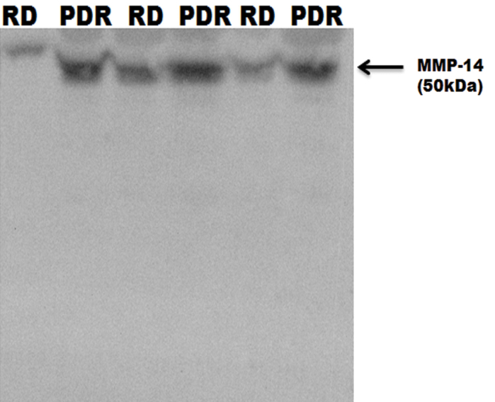
Methods: Vitreous samples from 34 patients with PDR and 18 nondiabetic patients and epiretinal membranes from 13 patients with PDR and the retinas of rats were studied with enzyme-linked immunosorbent assay, immunohistochemistry, western blotting, and real-time reverse transcription PCR (RT-PCR).

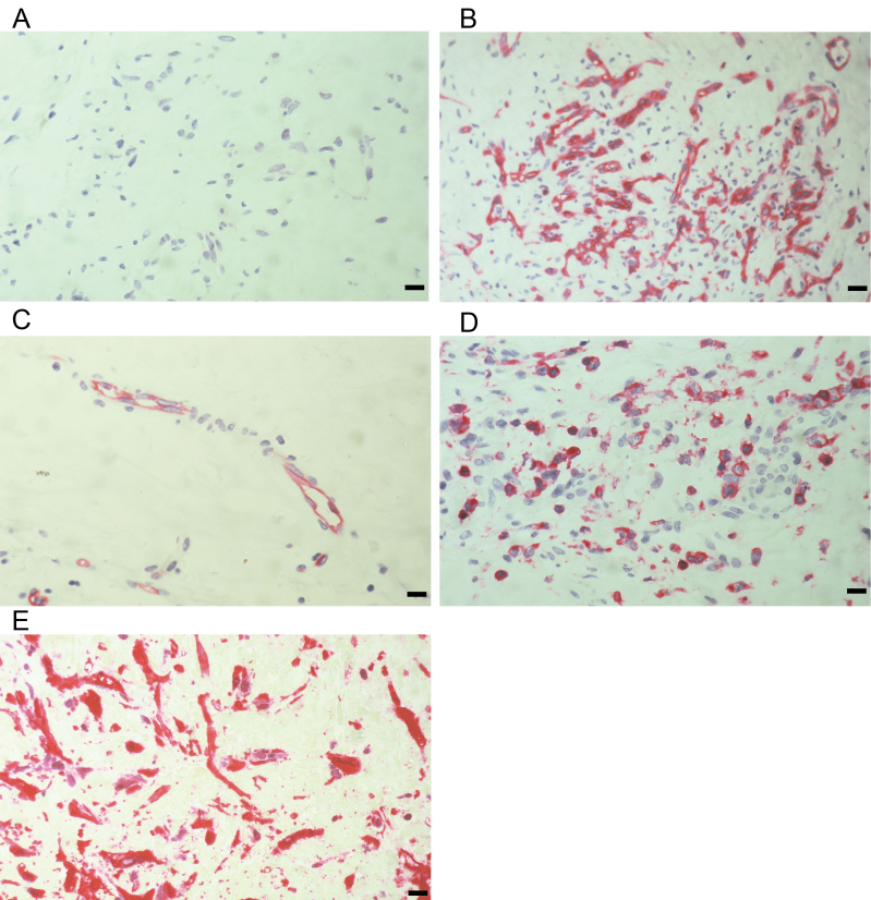
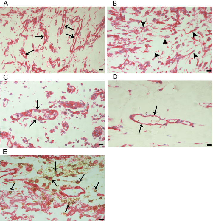
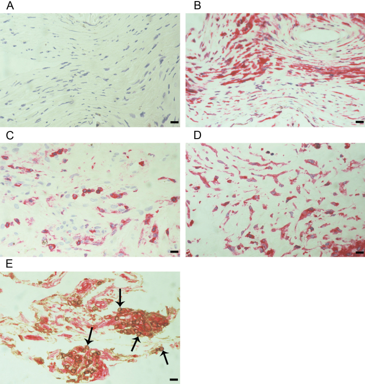
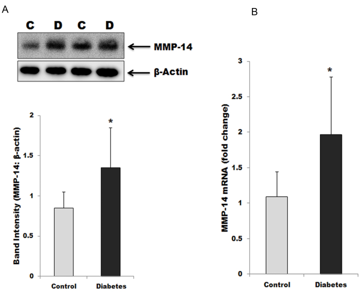
Results: The MMP-14, VEGF, and MMP-9 levels were statistically significantly higher in the vitreous samples from patients with PDR than in the samples from the nondiabetic controls (p<0.001 for all comparisons). The MMP-14 levels in patients with PDR with active neovascularization were statistically significantly higher than those in patients with inactive PDR (p<0.001). There were statistically significant positive correlations between levels of MMP-14 and levels of VEGF (r = 0.3; p = 0.032) and MMP-9 (r = 0.54; p<0.001). In the epiretinal membranes, MMP-14 was expressed in vascular endothelial cells, leukocytes, and myofibroblasts. Statistically significant positive correlations were detected between the numbers of blood vessels expressing CD31 and the numbers of blood vessels (r = 0.74; p = 0.004) and stromal cells (r = 0.72; p = 0.005) expressing MMP-14. Statistically significant increases of MMP-14 mRNA and protein were detected in rat retinas after induction of diabetes.

Conclusions: These results suggest that MMP-14 is involved in PDR angiogenesis.


Figures

Figure 1
Figure 1
The expression of matrix metalloproteinase-14 (MMP-14) in equal volumes (15 µl) of vitreous fluid samples obtained from patients with proliferative diabetic retinopathy (PDR) and from control patients with rhegmatogenous retinal detachment (RD) was determined with western blot analysis.
Figure 2
Figure 2
Proliferative diabetic retinopathy (PDR) fibrovascular epiretinal membranes. A: Negative control slide showing no labeling. Immunohistochemical staining for CD31 showing pathologic new blood vessels expressing this endothelial cell marker in (B) a membrane from a patient with active PDR and in (C) a membrane from a patient with inactive PDR. D: Immunohistochemical staining for CD45 showing numerous leukocytes in the stroma. E: Immunohistochemical staining for α-smooth muscle actin showing immunoreactivity in myofibroblasts (scale bar, 10 µm).
Figure 3
Figure 3
Proliferative diabetic retinopathy (PDR) fibrovascular epiretinal membranes. Immunohistochemical staining for matrix metalloproteinase-14 (MMP-14) showing immunoreactivity in the vascular endothelial cells (arrows), in intravascular leukocytes, in stromal cells, and in stromal spindle-shaped cells (arrowheads) in a membrane from a patient with active PDR (A, B, C) and in a membrane from a patient with inactive PDR (D). Notice that the membrane from the patient with inactive PDR is composed mostly of fibrous tissue. Double immunohistochemistry for CD45 (brown) and MMP-14 (red) in a membrane from a patient with active PDR demonstrated stromal cells coexpressing CD45 and MMP-14 (arrows; E). No counterstain to visualize the cell nuclei was applied (scale bar, 10 µm).
Figure 4
Figure 4
Proliferative vitreoretinopathy fibrocellular epiretinal membranes. A: Negative control slide showing no labeling. B: Immunohistochemical staining for α-smooth muscle actin showing immunoreactivity in spindle-shaped myofibroblasts. C: Immunoreactivity staining for CD45 showing immunoreactivity in leukocytes. D: Immunohistochemical staining for matrix metalloproteinase-14 (MMP-14) showing immunohistochemistry in spindle-shaped myofibroblasts. E: Double immunohistochemistry for CD45 (brown) and MMP-14 (red) demonstrates cells coexpressing CD45 and MMP-14 (arrows; scale bar, 10 µm).
Figure 5
Figure 5
Matrix metalloproteinase-14 (MMP-14) expression in diabetic rat retinas. A: MMP-14 expression was determined by western blot analysis on lysates of diabetic (D) and nondiabetic control (C) rats. After determination of the intensity of the MMP-14 protein band, intensities were adjusted to those of β-actin in each sample. Each measurement was performed at least three times. B: In addition, cell extracts were used to quantify the mRNA of MMP-14 by quantitative-RT-PCR. Results are expressed as mean ± standard deviation of 7-10 rats in each group. *The difference between the two means was statistically significant at 5% level of significance.

References

    1. Abu El-Asrar AM, De Hertogh G, Van den Eynde K, Alam K, Van Raemdonck K, Opdenakker G, Van Damme J, Geboes K, Struyf S. Myofibroblasts in proliferative diabetic retinopathy can originate from infiltrating fibrocytes and through endothelial-to-mesenchymal transition (EndoMT). Exp Eye Res. 2015;132:179–89. - PubMed
    1. Abu El-Asrar AM, Nawaz MI, De Hertogh G, Alam K, Siddiquei MM, Van den Eynde K, Mousa A, Mohammad G, Geboes K, Opdenakker G. S100A4 is upregulated in proliferative diabetic retinopathy and correlates with markers of angiogenesis and fibrogenesis. Mol Vis. 2014;20:1209–24. - PMC - PubMed
    1. Abu El-Asrar AM, Nawaz MI, Kangave D, Siddiquei MM, Geboes K. Angiogenic and vasculogenic factors in the vitreous from patients with proliferative diabetic retinopathy. J Diabetes Res. 2013;2013:539658. - PMC - PubMed
    1. El-Asrar AM, Nawaz MI, Kangave D, Geboes K, Ola MS, Ahmad S, Al-Shabrawey M. High-mobility group box-1and biomarkers of inflammation in the vitreous from patients with proliferative diabetic retinopathy. Mol Vis. 2011;17:1829–38. - PMC - PubMed
    1. Nawaz MI, Van Raemdonck K, Mohammad G, Kangave D, Van Damme J, Abu El-Asrar AM, Struyf S. Autocrine CCL2, CXCL4, CXCL9 and CXCL10 signal in retinal endothelial cells and are enhanced in diabetic retinopathy. Exp Eye Res. 2013;109:67–76. - PubMed

Publication types

MeSH terms